
|
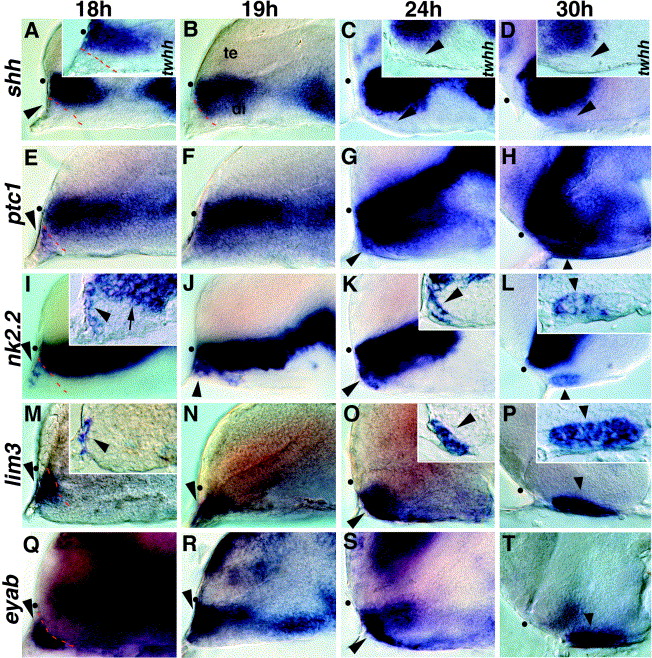
Fig. 4 Regionalization within the developing adenohypophysis. (A–D) Expression of shh and twhh (insets) is restricted to the neural tube; no expression is seen in anterior epidermal cells (arrowhead in A). (E–H) ptc1 expression extends into nonneural cells at the anterior edge of the embryo, anterior to the shh expression domain (arrowheads). (I, J) nk2.2 is expressed anterior to the neural tube at 18 h (arrowhead), and this expression is distinct from the neural expression domain (arrow in inset). (K, L) At 24 and 30 h, nk2.2 is expressed in the anterior region of the visibly thickened adenohypophyseal placode (arrowheads) with expression restricted to the anterior region at both ages (arrowheads in insets). (M, N) lim3 expression anterior to the neural tube at 18 and 19 h (arrowhead). (O, P) lim3 continues to be expressed in the entire thickened placodal region at 24 and 30 h (arrowheads). (Q–T) The placodal marker eyaB is expressed throughout the adenohypophyseal placode (arrowheads). All panels show lateral views, eyes removed, anterior to the left. Insets show parasagittal sections. te, telencephalon; di, diencephalon; dot shows position of optic recess.
Reprinted from Developmental Biology, 254(1), Sbrogna, J.L., Barresi, M.J., and Karlstrom, R.O., Multiple roles for Hedgehog signaling in zebrafish pituitary development, 19-35, Copyright (2003) with permission from Elsevier. Full text @ Dev. Biol.