
|
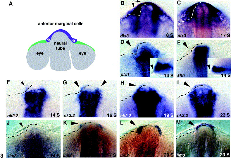
Fig. 3 ptc1 and nk2.2 expression precedes lim3 expression in preplacodal cells at the anterior margin of the neural tube. (A) Schematic of the zebrafish forebrain during somitogenesis, dorsal view, anterior up. Based on expression of lim3 and fate mapping experiments, the pituitary placode arises from medially located cells (purple) at the anterior edge of the neural tube Glasgow et al 1997 and Whitlock and Westerfield 2000. Asterisks show where the pituitary transcription factors lim3 and nk2.2 are first expressed. (B) At the 8-somite (S) stage, the early placodal marker dlx3 labels marginal cells spanning the midline of the neural tube and more lateral cells that coalesce to form the olfactory placodes (arrows). (C) At 17 S, dlx3 is expressed by a thinner band of preplacodal cells at the anterior margin. (D) The Hh receptor ptc1 is expressed in neural tissue and in anterior marginal cells (arrowheads) at 14 S. Inset shows lateral view. (E) shh expression is restricted to neural tissue, no expression is seen in epidermal cells (arrowheads). Inset shows lateral view. (F) nk2.2 expression begins anterior to the neural tube as early as 14 S and is often seen only on the left side (arrowhead). (G) By 16 S, nk2.2 is expressed anterior to the neural tube on both sides of the embryo (arrowheads). (H, I) Between 19 and 23 S, nk2.2 expression expands across the midline in anterior marginal cells (arrowheads). (J) At 14 S, no lim3 expression is detectable in anterior marginal cells (arrowhead). (K) lim3 expression is first detectable at approximately 17 S (arrowhead). (L, M) Between 20 and 23 S, lim3 expression expands across the midline (arrowheads). All panels show dorsal/anterior views of the anterior edge of the nervous system, anterior up. Insets in (D) and (E) show lateral views, anterior to the left. Dotted lines on left side of embryo show position of the visible border between neural tissue and epidermal cells at the anterior margin of the nervous system. This border was established by careful examination of magnified images and multiple labeled embryos.
Reprinted from Developmental Biology, 254(1), Sbrogna, J.L., Barresi, M.J., and Karlstrom, R.O., Multiple roles for Hedgehog signaling in zebrafish pituitary development, 19-35, Copyright (2003) with permission from Elsevier. Full text @ Dev. Biol.