
|
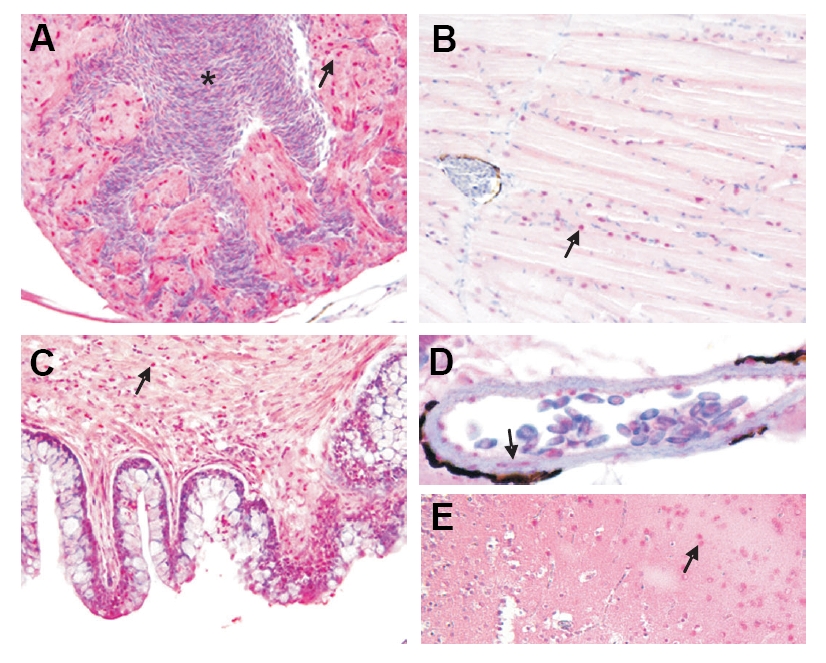
Fig. 4 Adult tissue expression of Srf1 protein. Adult (3 month) tissues were processed for immunohistochemistry as described in Materials and Methods. Arrows point to Srf1 nuclear staining (in red) in cardiomyocytes of the heart (A), skeletal muscle of posterior trunk (B), visceral SMC of gut (C), vascular SMC of dorsal aorta (D) and cortical neurons of brain (E). The asterisk in (A) indicates nucleated red blood cells within the ventricular chamber of the heart. Note the peri-aortic melanin (dark staining rim in panel D) that has recently been described and validated (Miano et al., 2006). Staining specificity was demonstrated with a competing peptide to human SRF (data not shown). Magnifications are 400x.