
|
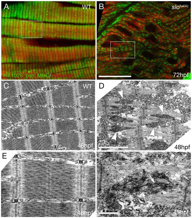
Fig. S2 slotθ44c mutants have a similar but slightly less severe muscle structure defect than sloθ45 mutants. (A,B) Muscle fibres double stained for primary sarcomeric components Actin and Myosin using phalloidin (red) and A4.1025 anti-MHC antibodies (green) in slotu44c mutants and siblings. slotu44c mutant fibres have some apparent organisation in both Actin and anti-MHC staining; however, compared to the sibling, the staining is disordered. Boxes indicate approximately similar fields of view shown in the electron micrographs shown in C,D. (C-F) Ultrastructural analysis of wild-type and slotu44c muscle fibres shows that the slotu44c fibres contain electron-dense bodies (arrowheads, D) located approximately between adjacent I-Z-I brushes. These bodies appear to be thickest in the region equidistant between adjacent Z-lines tapering away towards the Z-lines suggesting deformed A-bands. These electron-dense bodies were sometimes found to contain disordered thick-filament-like structures (arrows, F). Scale bars: 10 μm in A,B; 2 μm in C,D; 500 nm in E,F.