
|
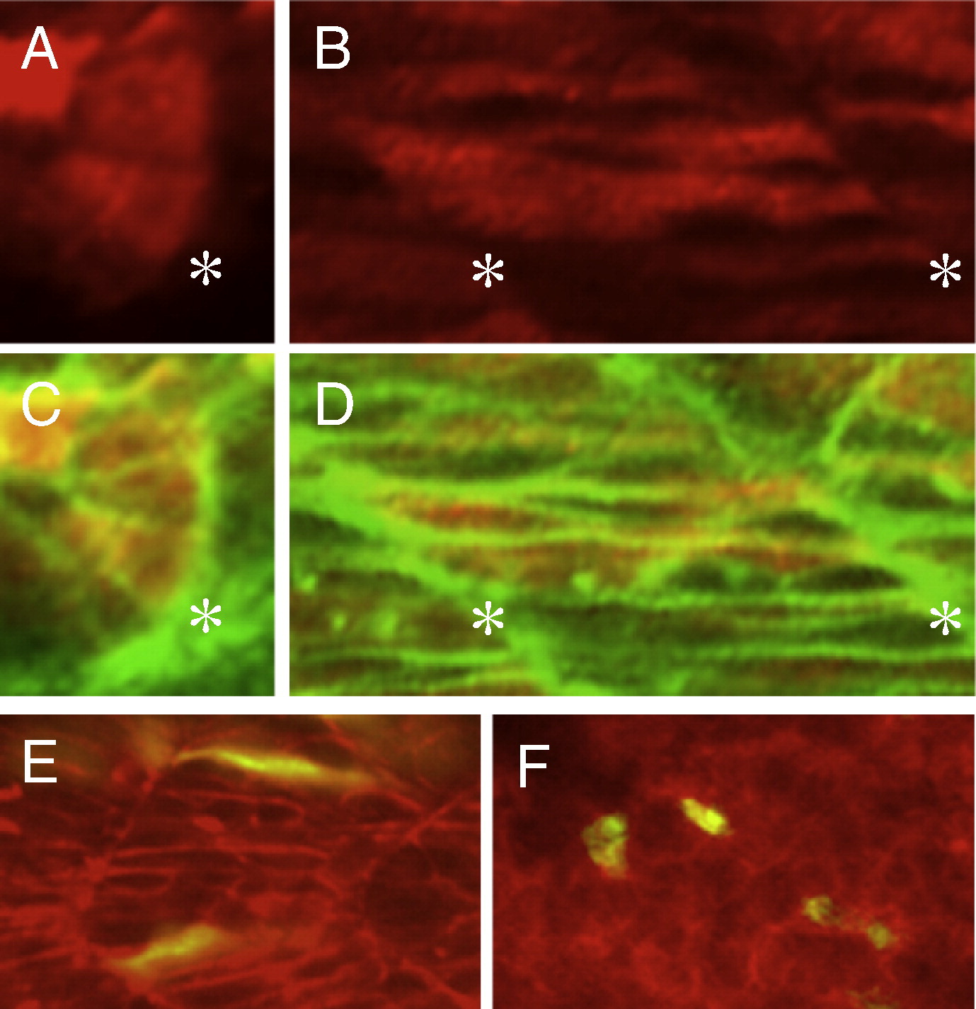
Fig. 8 Immunostaining of the Flag–Mys fusion protein and cell transpolantation. (A–D) Confocal images of embryos injected with flag–mys mRNA and stained with the anti-Flag antibody (red) and Phalloidin (green). (A, C) Dorsal views of the somite at 16 hpf. (B, D) Lateral views of the somite at 20 hpf. (C, D) Merged images. Asterisks indicate the positions of the somite boundaries. The Flag–Mys protein was distributed throughout the cytoplasm. (E, F) Confocal images of lateral views of transplanted embryos at 20 hpf. (E) Cells from embryos injected with ATG-MO, rhodamine-dextran and GFP mRNA were transplanted into a wild type embryo. (F) Cells from embryos injected with rhodamine-dextran and GFP mRNA were transplanted into an ATG-MO injected embryo. The recipient embryos were stained with phalloidin (red) and transplanted cells were labeled in yellow. The ATG-MO injected cells were elongated in the wild type embryo (E) and the wild type cells showed rounded shapes in the ATG-MO injected embryo.
Reprinted from Developmental Biology, 316(2), Kotani, T., and Kawakami, K., misty somites, a maternal effect gene identified by transposon-mediated insertional mutagenesis in zebrafish that is essential for the somite boundary maintenance, 383-396, Copyright (2008) with permission from Elsevier. Full text @ Dev. Biol.