
Fig. 1
- ID
- ZDB-IMAGE-080417-28
- Genes
- Publication
- Kotani et al., 2008 - misty somites, a maternal effect gene identified by transposon-mediated insertional mutagenesis in zebrafish that is essential for the somite boundary maintenance
- All Figures
- Figures for Kotani et al., 2008
|
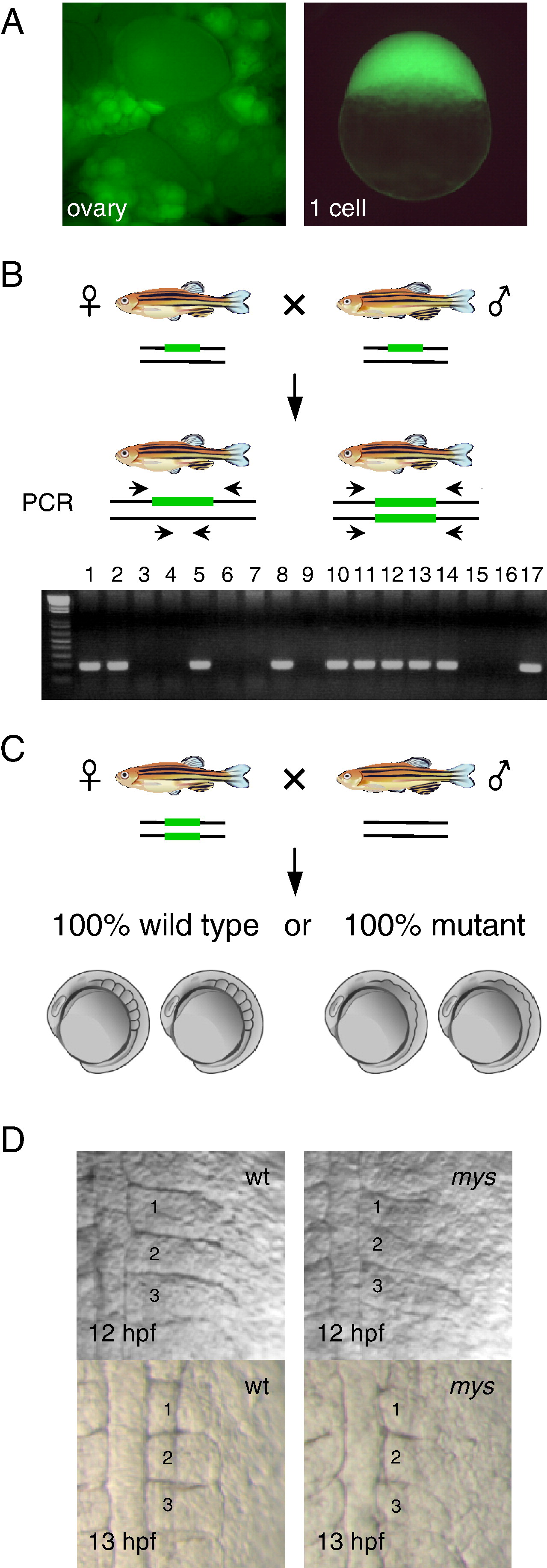
Fig. 1 A scheme for isolation of maternal effect mutants by transposon-mediated gene trapping. (A) Identification of embryos that express GFP at the one cell stage. GFP expression in the ovary (left) and in fertilized eggs (right) (SAG20A). (B) Identification of fish homozygous for an insertion. Heterozygous male and female fish are crossed and the progeny are analyzed by PCR using primers that locate both sides of the insertion (green bars). The gel is an example of the analysis. A PCR product is amplified from heterozygous fish (lanes 1–2,5,8,10–14 and 17) but not from homozygous fish (lanes 3,4,6,7,9,15 and 16). (C) Homozygous female fish is crossed with wild type male fish and their progeny are analyzed for developmental defects. (D) The somite boundary phenotype at the 3- and 6-somite stages (12 hpf and 13 hpf). (left) Wild type embryos. (right) Embryos from SAG20A homozygous female fish. Numbers show the somite numbers.
Reprinted from Developmental Biology, 316(2), Kotani, T., and Kawakami, K., misty somites, a maternal effect gene identified by transposon-mediated insertional mutagenesis in zebrafish that is essential for the somite boundary maintenance, 383-396, Copyright (2008) with permission from Elsevier. Full text @ Dev. Biol.