
|
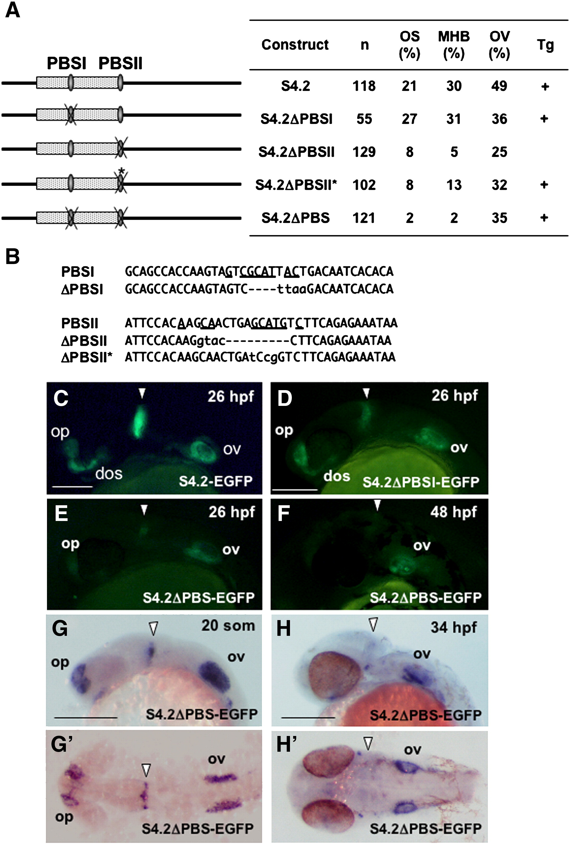
Fig. 8 Essential role of the two Pax2 binding sites in the regulatory function of the S4.2 enhancer. (A) Disruption of the Pax2 binding sites (PBSI and PBSII) in S4.2-EGFP. The percentages of injected embryos expressing GFP in the optic stalk (OS), MHB, and the otic vesicle (OV) at 24–26 hpf relative to the numbers of GFP-positive embryos (n) are shown on the right. The constructs for which expression was confirmed by stable expression in Tg embryos are marked with (+). (B) Mutations introduced into the two Pax2-biniding sites of S4.2-EGFP. Deletions and base substitutions are indicated by hyphens and lowercase letters, respectively. In case of ΔPBSI and ΔPBSII, an EcoRI and a KpnI sequence were introduced into the Pax2-binding core sites by inverse PCR, respectively, resulting in base deletion and substitution. Meanwhile, the Pax2 site was disrupted by site-directed mutagenesis for ΔPBSII*. (C–F) Stable GFP expression in Tg embryos harboring the intact (C) or mutated S4.2-EGFP constructs (D–F) was observed at 26 hpf (C–E) and 48 hpf (F). (G, H, G′, H′) Expression of egfp mRNA in the head of an S4.2ΔPBS-EGFP Tg embryo. Expression of S4.2ΔPBS-EGFP in the MHB was weak relative to that in the sensory regions at 19 hpf (20-somite stage)–26 hpf (E, G, G′), and it was disrupted later (F, H, H′). Lateral (C–H) and dorsal (G′, H′) views are shown with anterior to the left. Arrowheads indicate the MHB. dos, distal optic stalk; op, olfactory placode; ov, otic vesicle. Scale bars, 200 μm.
Reprinted from Developmental Biology, 316(2), Inoue, F., Parvin, M.S., and Yamasu, K., Transcription of fgf8 is regulated by activating and repressive cis-elements at the midbrain-hindbrain boundary in zebrafish embryos, 471-486, Copyright (2008) with permission from Elsevier. Full text @ Dev. Biol.