
|
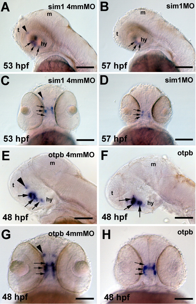
Fig. 5 sim1 and otpb are required for avt mRNA expression in the dorsal preoptic area. avt mRNA was detected by in situ hybridization histochemistry (ISH) in embryos that were injected with morpholino oligonucleotides (MO). A,C: Embryos injected with 0.5 ng of a sim1 four-base mismatch control MO (sim1 4mmMO). B,D: Embryos injected with 0.5 ng of sim1 MO. E,G: Embryos injected with 0.5 ng of an otpb four-base mismatch control MO (otpb 4mmMO). F,H: Embryos injected with 0.5 ng of otbp MO. A,B,E,F: Lateral views, dorsal is up, anterior to the left. C,D,G,H: Ventral views, anterior is up. The ages of the embryos are indicated in hours post fertilization (hpf). The avt-expressing cell groups in the ventral hypothalamus are indicated by black arrows, while the avt cell group in the dorsal preoptic area is indicated with a black arrowhead. E,F: The eyes have been removed. A brown shadow from the eye lens is present in A, but the avt-expressing cells are stained dark blue and are indicated with arrows. hy, hypothalamus; m, midbrain; t, telencephalon. Scale bars = 100 μm.