
|
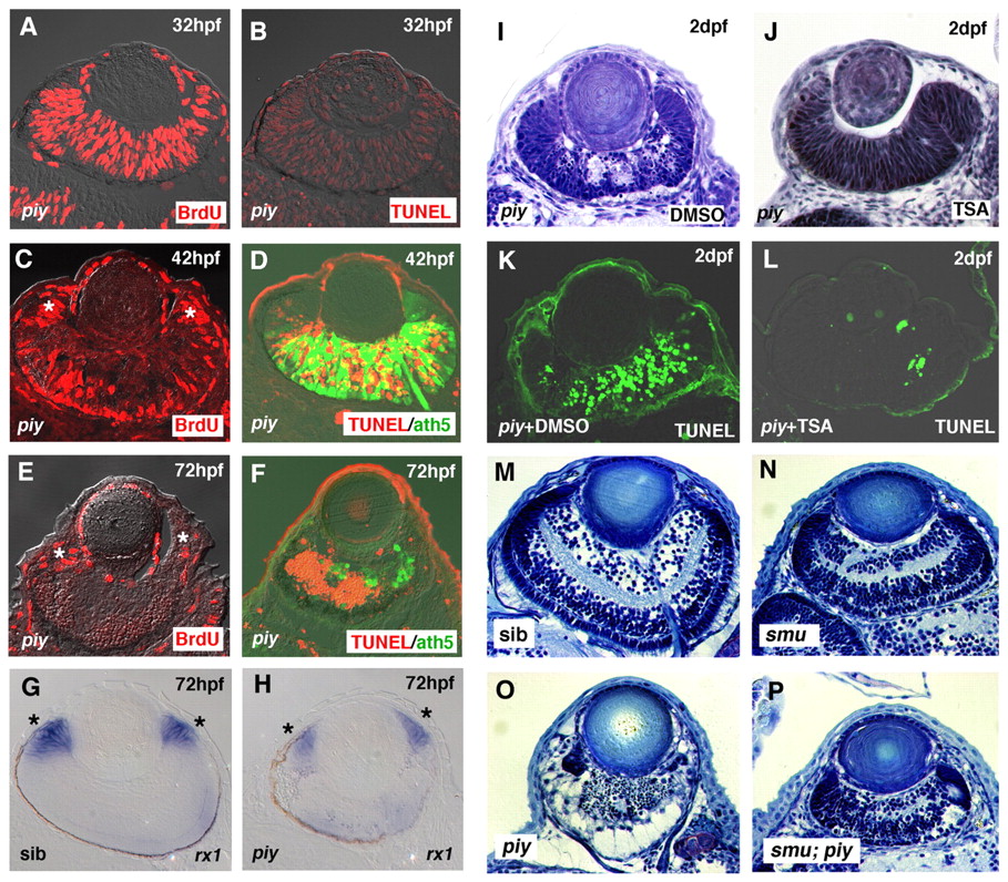
Fig. 2 Apoptosis occurs in differentiating neurons in the piy mutant retina. (A-F) BrdU labeling (A,C,E) and TUNEL (B,D,F) of piy mutant zebrafish retinas. At 32 hpf, many retinal cells incorporate BrdU (A, red) and there are no TUNEL-positive cells (B, red). At 42 hpf, many neurons are produced in the central retina and these express the neuronal marker ath5 (D, green). Apoptotic cells are observed in the ath5-positive central retina, but are rare in the CMZ (D, red). At 72 hpf, dense dead cells are observed in the central retina (F, red) and ath5-positive neurons markedly decrease in number (F, green), suggesting that differentiating neurons undergo apoptosis in the piy mutant. Cells in the CMZ incorporate BrdU (C,E, asterisks), suggesting that retinal stem cells continue to proliferate in the piy mutant. (G,H) In situ hybridization of wild-type (G) and piy mutant (H) retinas with rx1 RNA probe (asterisks). (I,J) Plastic sections of piy mutant retinas treated with DMSO (I) or TSA (J). (K,L) TUNEL (green) of piy mutant retinas treated with DMSO (K) or TSA (L). (M-P) Plastic sections of wild-type (M), smu (N), piy (O) and smu; piy (P) mutant retinas. Although retinal neurogenesis is delayed in the smu mutant, retinal lamination occurs until 3 dpf (N). In the smu; piy double mutant, retinal cell apoptosis is suppressed (P).