
Image
|
Figure Caption
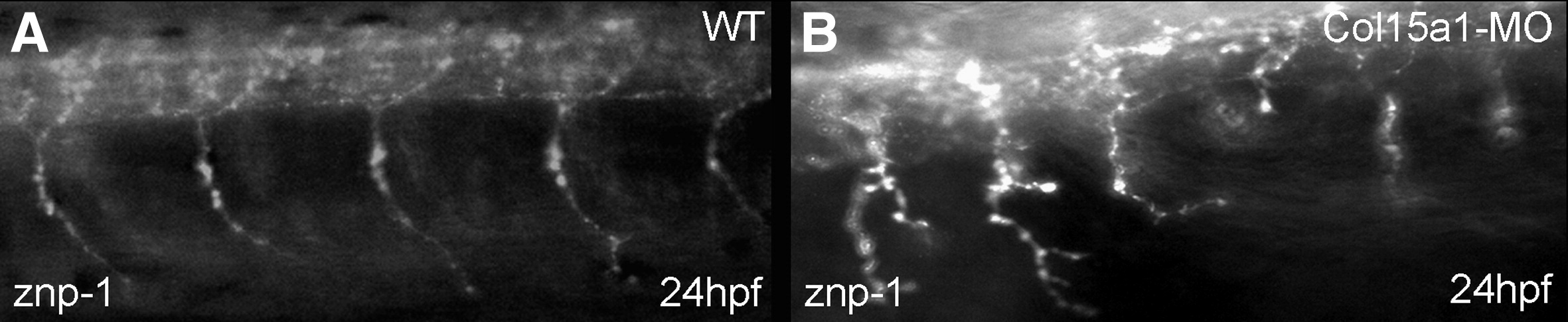
Fig. 11 Projection pattern of primary CaP and MiP motorneurons in the wild-type (A) and Col15a1-MO (B) embryos. (A, B) Lateral views of confocal scans of 24 hpf whole mount embryos stained with znp-1 antibody. Ventral motor nerve extends shorter or forms extended branched axons in B. Magnification: A, B: x 400.
Figure Data
Acknowledgments
This image is the copyrighted work of the attributed author or publisher, and
ZFIN has permission only to display this image to its users.
Additional permissions should be obtained from the applicable author or publisher of the image.
Reprinted from Developmental Biology, 316(1), Pagnon-Minot, A., Malbouyres, M., Haftek-Terreau, Z., Kim, H.R., Sasaki, T., Thisse, C., Thisse, B., Ingham, P.W., Ruggiero, F., and Le Guellec, D., Collagen XV, a novel factor in zebrafish notochord differentiation and muscle development, 21-35, Copyright (2008) with permission from Elsevier. Full text @ Dev. Biol.