
|
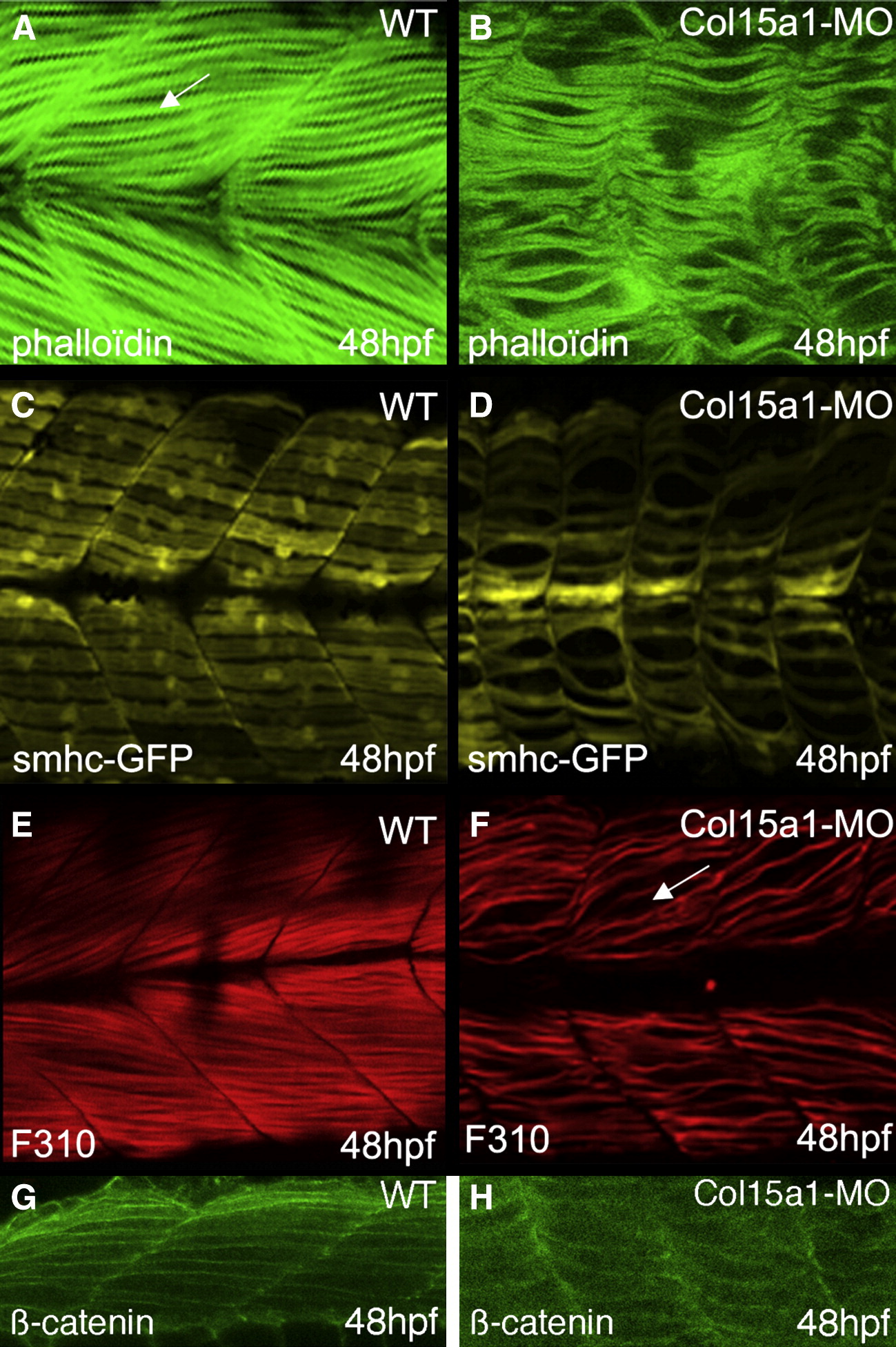
Fig. 8 The lack of collagen XV disrupts slow and fast muscle patterning. (A, B) Lateral views of the myotomal segments of 48 hpf wild-type (A) and Col15a1-MO (B) embryos, revealing the slow and fast fibers stained with phalloïdin-FITC (arrows). (B) Similar views of morphants reveal an important disorganization of myofibrils. (C) 48 hpf wild-type and (D) Col15a1-MO smhc-GFP transgenic embryos, showing the organization of the slow fibers. Note that the slow fibers are collapsed in the Col15a1-MO embryos and that the nuclei are less visible. (E, F) Medial views of wild-type (E) and Col15a1-MO (F) embryos, revealing the fast fibers stained with mAb F310. At 48 hpf, only few Col15a1-MO fast fibers are observed, and large areas of the muscle are not stained (arrows). (G, H) Lateral views of 48 hpf wild-type (G) and Col15a1-MO (H) staining with β-catenin antibodies show that the lack of collagen XV does not lead to a decrease in muscle cell number. Magnification: A–H: x 300.
Reprinted from Developmental Biology, 316(1), Pagnon-Minot, A., Malbouyres, M., Haftek-Terreau, Z., Kim, H.R., Sasaki, T., Thisse, C., Thisse, B., Ingham, P.W., Ruggiero, F., and Le Guellec, D., Collagen XV, a novel factor in zebrafish notochord differentiation and muscle development, 21-35, Copyright (2008) with permission from Elsevier. Full text @ Dev. Biol.