
|
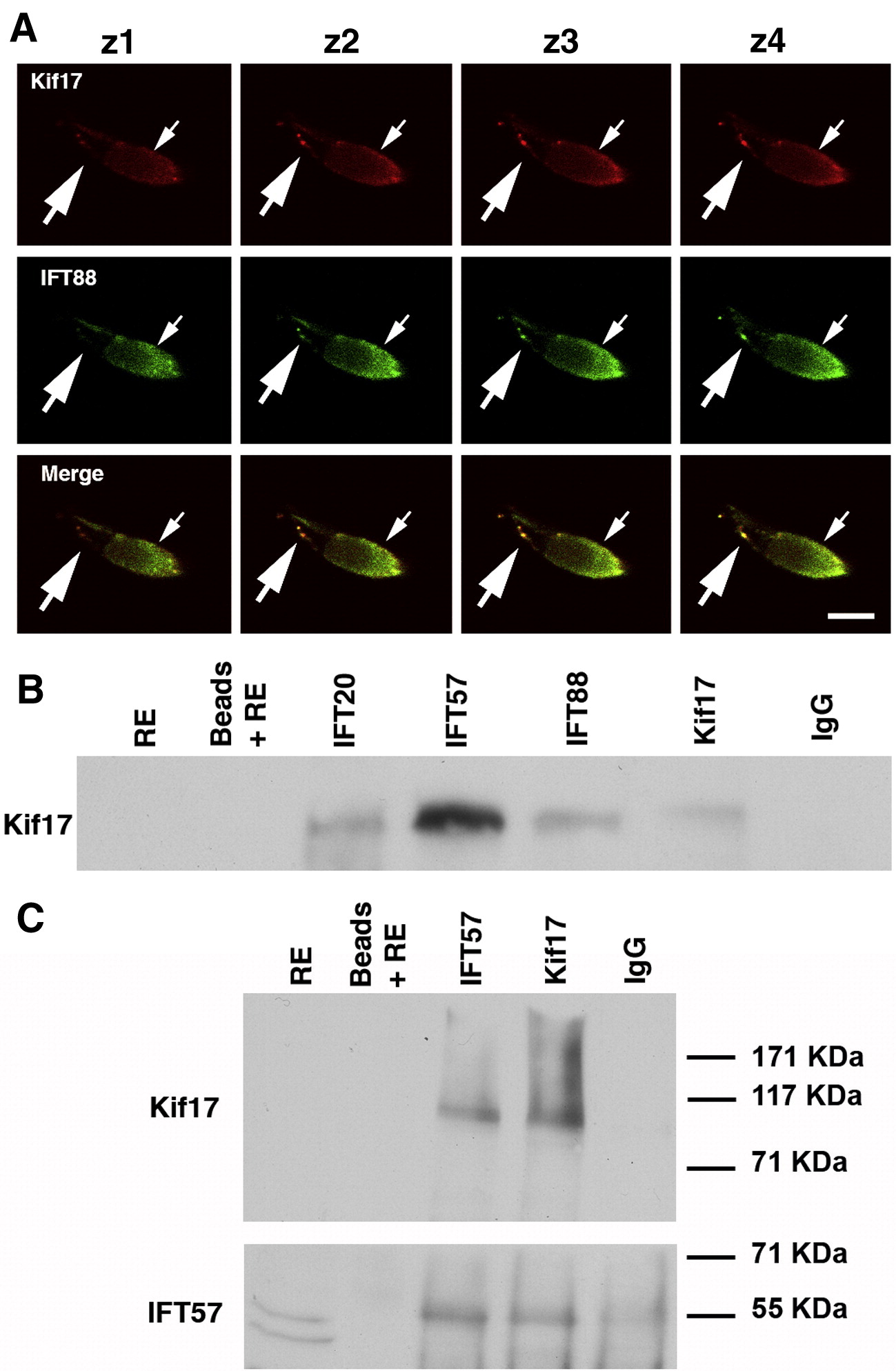
Fig. 3 Association of Kif17 with IFT complex proteins. (A) Consecutive confocal Z-slices of a cone double labeled with Kif17 and IFT88 antibodies. The small arrow indicates the position of the ellipsoid and the large arrow points to punctate staining along the axoneme. Bar = 10 μm. (B) Co-immunoprecipitation of Kif17 with antibodies to IFT20, IFT57, IFT88, and Kif17. IP antibodies are shown at the top and western blot antibodies on the left. Kif17 was not detected in the RE, which was at 1/5 of the volume used for IP; Kif17 was readily detected when the extract load was higher (see Fig. 1A). Beads plus RE without antibody and a mixture of IgG1 and IgG2 were used as controls for non-specific binding. In separate gels, IFT antibodies were shown to co-IP using anti-IFT antibodies in zebrafish as previously shown for mouse retina (Baker et al., 2003). (C) IP experiment as in panel B in which the blot was cut into two pieces; the upper portion was stained for Kif17 and lower portion for IFT57. Note that as reported in mice (Pazour et al., 2002), two bands are detected with the IFT57 antibody but only the upper band is present in the co-IPs.
Reprinted from Developmental Biology, 316(1), Insinna, C., Pathak, N., Perkins, B., Drummond, I., and Besharse, J.C., The homodimeric kinesin, Kif17, is essential for vertebrate photoreceptor sensory outer segment development, 160-170, Copyright (2008) with permission from Elsevier. Full text @ Dev. Biol.