
|
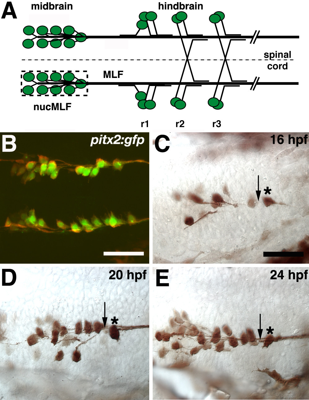
Fig. 1 Initial extension and convergence of MLF axons.(a-e) Ventral views, anterior to the left. (a) Schematic representation of MLF axons and hindbrain axons that grow along the MLF. The dashed line denotes the ventral midline and the dashed box surrounds the 'nucMLF zone'. r = rhombomere. (b) Confocal projection of 20 hpf Tg(pitx2c:gfp) embryo stained with anti-GFP (green) and ZN-12 antibodies (red). (c-e) Whole mount preparations of Tg(pitx2c:gfp) embryos stained with anti-GFP at 16 (c), 20 (d), and 24 (e) hpf. Midline is up. Asterisks denote the caudal-most nucMLF cell and arrows indicate the 'convergence point'. Scale bar = 25 μm.