
Image
|
Figure Caption
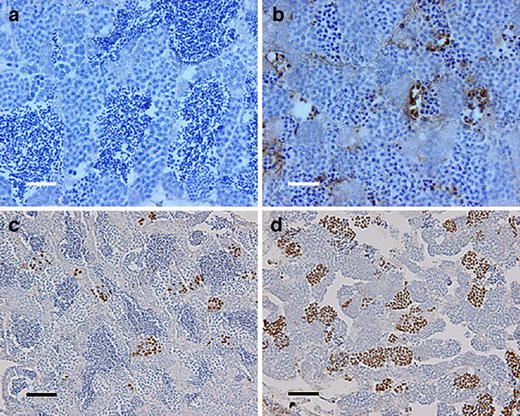
Fig. 2 Testis immunohistochemistry of mlh -/- zebrafish. a, b TUNEL staining of mlh1 +/+ and mlh1 -/- fish, respectively. Large numbers of apoptotic cells can be seen in mutant testis (brown). Wild-type testis exhibits a low incidence of apoptosis. c, d Histone H3 staining of cells in metaphase from mlh1 +/+ and mlh1 -/- fish, respectively. Note the higher incidence of stained cells in mutant testis indicating a higher number of mitotic and meiotic cells in metaphase. Bars 25 &mum (a, b), 50 μm (c, d)
Figure Data
Acknowledgments
This image is the copyrighted work of the attributed author or publisher, and
ZFIN has permission only to display this image to its users.
Additional permissions should be obtained from the applicable author or publisher of the image.
open access
Full text @ Cell Tissue Res.