
Image
|
Figure Caption
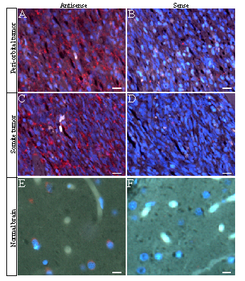
Fig. 10 The GFAP expression in primary tumors developed in the zebrafish p53M214K mutant line. Paraffin sections were hybridized with GFAP antisense (A, C, and E) and sense riboprobes (B, D, and F). Expression of GFAP was observed in a periorbital tumor (A, red) and a somite-derived tumor (B, red), compared with the normal levels of GFAP expression in normal brain cortex as a positive control (E, red). Blue indicates nuclear DNA staining with DAPI. (Bar, 40x)
Figure Data
Acknowledgments
This image is the copyrighted work of the attributed author or publisher, and
ZFIN has permission only to display this image to its users.
Additional permissions should be obtained from the applicable author or publisher of the image.
Full text @ Proc. Natl. Acad. Sci. USA