
IMAGE
Fig. S2
- ID
- ZDB-IMAGE-080401-21
- Genes
- Publication
- Godinho et al., 2007 - Nonapical symmetric divisions underlie horizontal cell layer formation in the developing retina in vivo
- All Figures
- Figures for Godinho et al., 2007
Image
|
Figure Caption
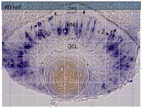
Fig. S2 Cross-section of the eye after in situ hybridization using a ptf1a antisense probe (see Lin et al., Dev. Biol. 274, 491-503 (2004) for Methods). Arrows, location of outer plexiform layer; ONL, outer nuclear layer; INL, inner nuclear layer; GCL, ganglion cell layer. Asterisk: labeled cell in amacrine cell layer. (1,2) Labeled cells in outer half of INL.
Figure Data
Acknowledgments
This image is the copyrighted work of the attributed author or publisher, and
ZFIN has permission only to display this image to its users.
Additional permissions should be obtained from the applicable author or publisher of the image.
Full text @ Neuron