
|
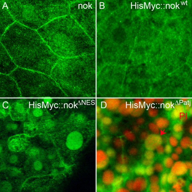
Fig. 2 The nok NES motif is required for nuclear export. Images represent reconstructions of confocal Z-stack sections imaged on late gastrula stage whole mounts. A: Endogenous nok mostly localizes to the outer cell membrane of EVL cells whereas nuclear localization is barely detectable. B: HisMyc-tagged nokwt fusion protein detected with anti-Myc antibody, green. The wt protein localizes to the outer cell membrane in EVL cells. C: HisMyc-tagged nokΔNES (green) localizes to the nucleus of EVL cells and lower levels of protein are also present at the outer cell membrane. D: HisMyc-tagged nokΔPatj localizes to the nucleus and lower levels are present at the outer cell membrane (green). Red arrow indicates an EVL cell nucleus, which is large compared with the smaller DL cell nuclei located just underneath. Propidium iodide (PI) nuclear counterstain in red.