
Image
|
Figure Caption
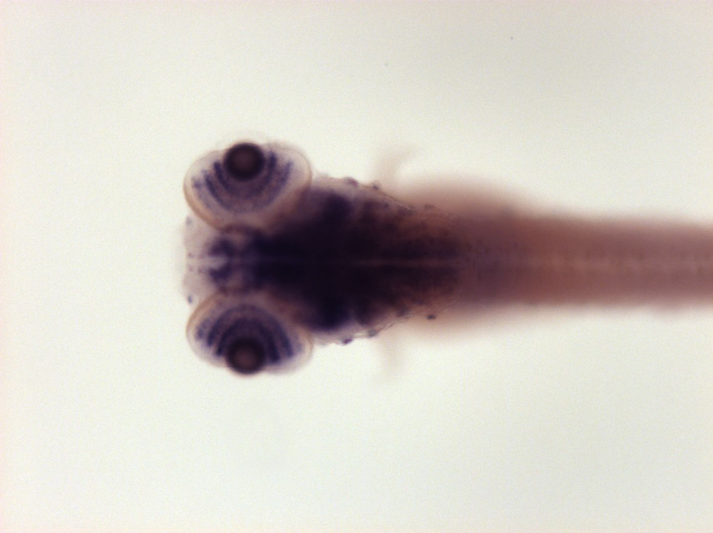
Fig. 7
brain marginal zone, ganglion and inner cell layer of retina
Please note that in 5 day old embryos some structures are not accessible to the probe (such as notochord, most of the trunk and tail). Therefore the description of the expression pattern is only partial.
Orientation
| Preparation | Image Form | View | Direction |
| whole-mount | still | dorsal | anterior to left |
Figure Data