
Fig. 7
- ID
- ZDB-IMAGE-080326-87
- Genes
- Publication
- Sato-Maeda et al., 2008 - Position fine-tuning of caudal primary motoneurons in the zebrafish spinal cord
- All Figures
- Figures for Sato-Maeda et al., 2008
|
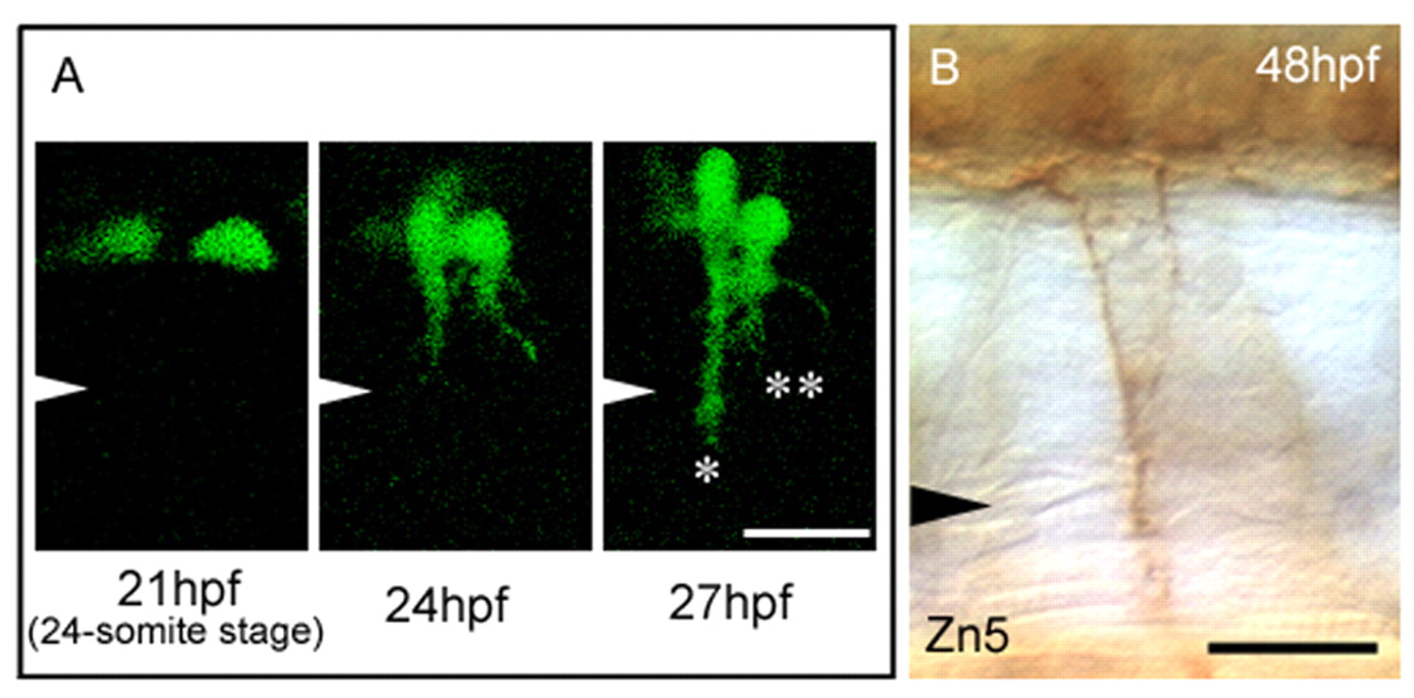
Fig. 7 Abnormal CaP cell positioning results in double exit points in both primary motoneurons (PMNs) and secondary motoneurons (SMNs). (A) Sequential images at the 18th somite level in an nrp1a:gfp transgenic embryo with Nrp1a-knockdown. Two separated CaP cell bodies (21 hpf) extended their axons to form double exit points (24 hpf). At 27 hpf, one axon extended beyond the horizontal myoseptal level (*), but the other axon did not (**). Arrowheads indicate the level of the horizontal myoseptum. (B) The double exit phenotype of SMN axons was observed at the same position as in A at 48 hpf. SMN axons were immunostained with mAb Zn5. Scale bars: 20 μm.