
|
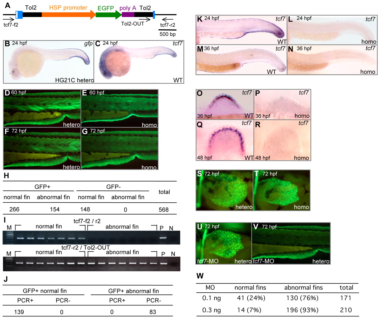
Fig. 3 The HG21C insertion disrupted the tcf7 gene. (A) The structure of the T2KHG insertion in the tcf7 gene. T2KHG is composed of Tol2 sequences (black), the zebrafish hsp70 promoter (orange), the GFP gene (green) and the SV40 polyA signal (purple). Exons (blue boxes) and 5′ UTR (white box) of the tcf7 gene are shown. Black arrows indicate positions and directions of primers. (B) Whole-mount in situ hybridization of an HG21C heterozygous embryo at 24 hpf using the gfp probe. (C) Whole-mount in situ hybridization of a wild-type embryo at 24 hpf using the tcf7 probe. (D-G) The median fin fold of HG21C embryos. Heterozygous (D,F) and homozygous (E,G) embryos at 60 hpf (D,E) and 72 hpf (F,G). (H) Summary of the linkage between the fin phenotype and the GFP expression. (I) Examples of the genotype analysis by PCR using tcf7-f2 and tcf7-r2 (top) and control PCR using tcf7-r2 and Tol2-OUT (bottom). M, DNA size marker; N, no DNA; P, positive control. (J) Summary of the genotype analysis. (K-R) Whole-mount in situ hybridization using the tcf7 probe. Wild-type embryos at 24 hpf (K) and 36 hpf (M), and HG21C homozygous embryos at 24 hpf (L) and 36 hpf (N). The pectoral fin buds of wild-type embryos at 36 hpf (O) and 48 hpf (Q), and HG21C homozygous embryos at 36 hpf (P) and 48 hpf (R). (S,T) The pectoral fins of 72 hpf heterozygous (S) and homozygous (T) HG21C embryos. (U,V) Microinjection of tcf7-MO. The pectoral fin (U) and the median fin (V) of MO-injected HG21 heterozygous embryos at 72 hpf. (W) Summary of the numbers of MO-injected embryos that showed abnormal median fin folds and pectoral fins.