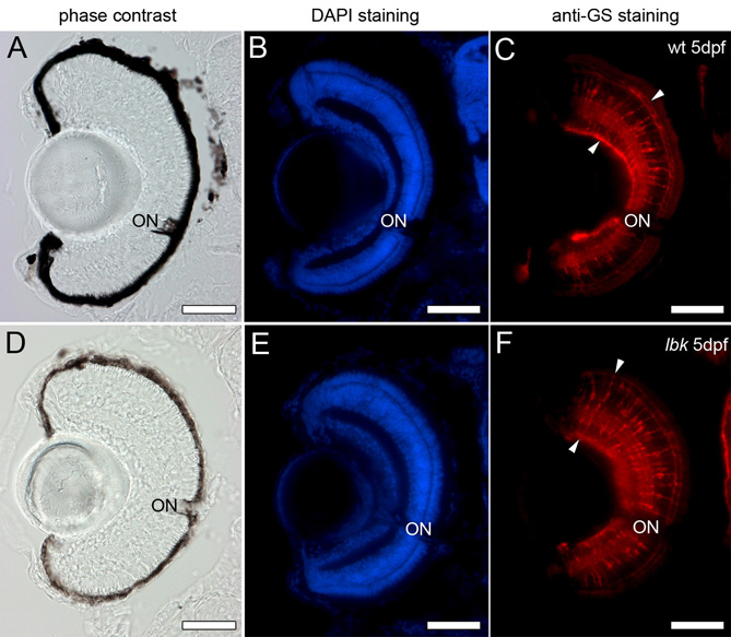
Fig. S6 Retinal Müller glia cells show a normal morphology in lbk mutants. Cryosections of 5 dpf wild-type and lbk eyes were stained with DAPI (blue; panels B and E) to show the position of the nuclei in the retina and an antibody against the glia-specific enzyme glutamine synthetase to label the retinal Müller glia cells (red; arrowheads in C and F). Phase-contrast images of the sections of wild-type and lbk eyes are shown in A and D, respectively. No obvious differences in the number, arrangement and morphology of the Müller glia cells were observed between wild-type and lbk larvae. ON, optic nerve. Scale bars: 50 μm.
Image
Figure Caption
Figure Data
Acknowledgments
This image is the copyrighted work of the attributed author or publisher, and
ZFIN has permission only to display this image to its users.
Additional permissions should be obtained from the applicable author or publisher of the image.
Full text @ Development

