
Image
|
Figure Caption
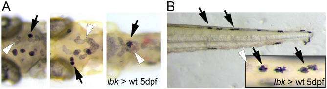
Fig. S4 Transplantation experiments show that the defect in lbk is cell-autonomous. Bright-field images of the dorsal head region (A) and tail (B) of four dark-adapted 5 dpf larvae. The skin melanocytes that have developed into normally pigmented melanocytes in an lbk-/- mutant background (arrows). Inset in B is a higher magnification dorsal view of four melanocytes with wild-type appearance indicated by arrows in B. Melanocytes displaying the severe hypopigmentation characteristic of lbk (white arrowheads) can be seen next to cells showing wild-type levels of melanin (black arrows).
Acknowledgments
This image is the copyrighted work of the attributed author or publisher, and
ZFIN has permission only to display this image to its users.
Additional permissions should be obtained from the applicable author or publisher of the image.
Full text @ Development