
|
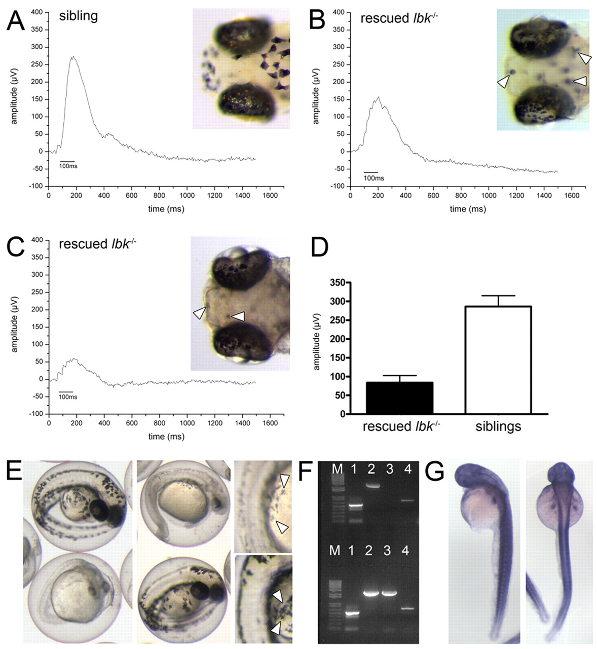
Fig. 6 The lbk phenotype can be partially rescued by the expression of vam6 in lbk-/- larvae and phenocopied by vam6 knock-down. (A-C) Electroretinogram analyses of 5 dpf vam6 vector-injected and heat-shocked sibling (A) and lbk-/- (B,C) larvae. In contrast to uninjected lbk larvae (Fig. 3B), injected lbk larvae displayed significant b-waves, indicating partial rescue of retinal function. The b-wave amplitude varied between different lbk larvae with some showing up to 60% of the wild-type b-wave amplitude (B), while others reach only ∼20% (C). Insets show head regions of vam6 vector-injected sibling (A) and lbk larvae (B,C) and the extent of rescue in RPE and skin melanocyte pigmentation. Arrowheads indicate skin melanocytes with near wild-type levels of melanin in rescued larvae. (D) Quantification of the observed ERG rescue: sibling larvae display an average b-wave amplitude of 286 μV, vam6 vector-injected lbk larvae show an average b-wave amplitude of 83 μV (n=7 for both sets of larvae; error bars show standard deviations). (E) Phenotype of the vam6 knock-down at 36 hpf showing the hypopigmentation of the RPE and skin melanocytes (insets; arrowheads) characteristic of lbk. However, the knock-down results in additional phenotypes not observed in lbk, including a small head and eyes and a shortened body axis. Larvae displaying wild-type levels of melanin are age-matched control morpholino-injected individuals. (F) PCR analysis for the presence of vam6 transcripts in RNA isolated from wild-type zebrafish zygotes (upper half of gel) and 24 hpf embryos (lower half). β-Actin (expressed at all developmental stages) and pmel17 [onset of expression: ∼20 hpf (Schonthaler et al., 2005)] served as controls. Lane M, 100 bp ladder; lane 1, amplification of a 530 bp β-actin cDNA fragment; lanes 2 and 4, amplification of two different fragments of the zebrafish vam6 cDNA (2628 bp and 707 bp) with independent primer pairs; lane 3, amplification of a 2538 bp fragment of the pmel17 cDNA. (G) Whole-mount in situ hybridisation on PTU-treated 48 hpf wild-type embryos (left, lateral view; right, dorsal view) showing vam6 expression.