
|
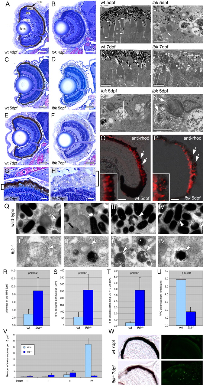
Fig. 2 lbk larvae display a severely compromised RPE and shorter PRC outer segments. (A-H) Sections of lbk (B,D,F,H) and sibling (A,C,E,G) eyes show that overall eye development proceeds normally in lbk, including the formation of a multi-layered neural retina. Beginning at 4 dpf (A,D), however, the retinal pigment epithelium (RPE) is severely hypopigmented and becomes progressively inflated. At 7 dpf (E-H), the RPE in lbk is threefold thicker than in age-matched siblings (G,H; brackets). The inner retina in lbk is morphologically indistinguishable from wild-type siblings. INL, inner nuclear layer; PRC, photoreceptor cells; RGC, retinal ganglion cells. (I-N) TEM sections reveal that the lbk RPE contains very few mature melanosomes. The majority of melanosomes are aberrantly shaped and show regions that lack pigment. Moreover, the mutant RPE is filled with numerous vesicles. The number of vesicles increases from 5-7 dpf (compare J with L). The microvilli of the RPE (I, arrowheads) that normally interdigitate with the outer segments of PRCs are significantly reduced in lbk (J; arrow). Furthermore, the outer segment length is reduced. Some lbk PRCs virtually lack outer segments (L, arrowheads). (M,N) Higher magnification of vesicles containing undigested PRC outer segments (arrows) in the lbk RPE (N shows a higher magnification of the boxed area in M). M, PRC mitochondria; N, PRC nucleus; OS, PRC outer segment. (O,P) Merged phase contrast and fluorescence images of eye sections stained for rhodopsin (red) show an increase of discrete rhodopsin-positive vesicles in the lbk RPE (P, arrows) versus continuous PRC OS in the wild type (O, arrows). Insets show higher magnifications of parts of the RPE from the sibling and mutant retinas shown in O and P. (Q) Images showing melanosomes at different stages of maturation (I-IV) in the sibling (top) and lbk (bottom) RPE at 5 dpf. Arrowheads indicate melanosomes of the respective stages. (R-U) Statistical analyses of RPE thickness (R), average RPE cell area per nucleus (S), average number of vesicles containing PRC outer segments (T) and PRC outer segment length (U) in sibling and lbk larvae at 5 dpf. Error bars indicate standard deviations; P values were calculated using Student′s t-test. (V) Quantification of melanosomes at different stages of maturation in the lbk and sibling RPE at 5 dpf. Counts represent the numbers of melanosomes in areas of 10 µm2. Although there are no substantial differences between the numbers of stage I-III melanosomes, mutant larvae contain significantly fewer mature (stage IV) melanosomes. (W) TUNEL labelling of cryosections shows increased levels of apoptosis in lbk (bottom right). The relatively dark appearance of the mutant RPE results from the thickness of these sections (40 μm). All images were derived from mid-transversal sections of the eye. Scale bars: 50 μm in A-F; 10 μm in G,H; 5 μm in I-M; 2.5 μm in N; 25 μm in O,P.