
|
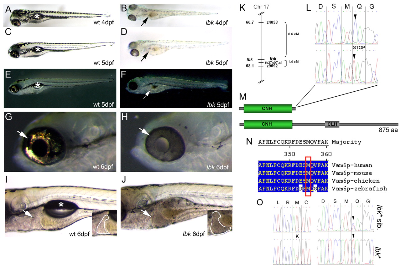
Fig. 1 A mutation in vam6 causes hypopigmentation and hepatomegaly in lbk. (A-D) Bright-field images show the hypopigmentation of skin melanocytes and the RPE, and a brownish discolouration of the intestinal tract (B,D, arrows) in lbk. Asterisks in A,C indicate the swim bladders in wild-type larva. (E-H) Dark-field images show an absence of iridophore reflections in the lbk skin and eye (G,H; arrows). Note the translucency of the lbk RPE in H. Arrow in F indicates the intestinal tract. Asterisk in E indicates the lac of swim bladder. (I,J) lbk larvae display an enlarged liver (insets in I,J; outlined areas) and lack swim bladders (asterisk). (K-O) The gene for Vam6p is mutated in lbk. (K) Genetic mapping of lbk resulted in linkage to SSLP markers z9692 and z4053 on chromosome 17. RH mapping of zebrafish vam6 resulted in linkage to EST fc27c07.x1, which was found to be located close to z9692 on the T51 RH map and thus served as an anchor to link the two maps. (L) Sequencing of cDNA from lbk-/- and homozygous wild-type larvae revealed a nucleotide exchange at position 1066 from C→T (arrowheads), resulting in a premature STOP codon. (M) Domain structures of the wild-type Vam6p (bottom) and the protein truncated in lbk lacking the C-terminal 520 amino acids (top), including the clathrin homology (CLH) repeat domain and Ypt7p-interacting sequences. (N) Sequence alignment of human, mouse, chicken and zebrafish Vam6p proteins shows the high level of conservation in the region affected by the mutation (red box). (O) Sequencing of the complete vam6 cds from the lbk* mutant revealed that this larva was heterozygous at two positions: the C→T nucleotide exchange identified in lbk (right panel, lower trace; arrowhead) and a novel T→A exchange at position 374 of the vam6 cds (left panel, lower trace). The latter results in the exchange of a conserved methionine (M) at amino acid position 125 for a lysine (K) in the N-terminal citron homology (CNH) domain (see Figs S2, S3 in the supplementary material). Sequencing of the complete vam6 cds from lbk* siblings showed the heterozygous presence of the lbk mutation (right panel, upper trace; arrowhead), but the absence of the T→A exchange at position 374 (left panel, upper trace).