
|
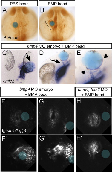
Fig. 7 BMP Beads Direct Cardiac Jogging (A and B) Phospho-Smad 1,5,8 antibody staining after implantation at the 15-somite stage of a PBS bead (A) or a BMP bead (B). Bracket in (B) indicates the induced phospho-Smad staining in close proximity of the BMP bead. Dorsal view with anterior to the top of 25-somite stage embryos. (C–E) ISH with antisense cmlc2 riboprobe of embryos injected with a bmp4 MO and a BMP bead implanted at the 15–18-somite stage. (C) The heart tube is redirected to a BMP bead implanted on the right side (11/15), dorsal view with anterior to the top, arrow indicates position of the BMP bead. (D) In a lateral view, arrows indicate CPCs that have been recruited toward the BMP bead (n = 7/8). Arrowheads (E) point to a layer of CPCs that form a layer on the bead's surface. (E) is an enlargement of (D). (F–H′) Selected images of a confocal time-lapse recording of bmp4 morphants (F–G′) or a bmp4 has2 double morphant (H and H′) with a BMP bead placed on the right side. Images represent the start (F–H) and end point (F′,G′,H′) of the time-lapse of the three individual embryos. Position of the BMP beads is marked by the blue circle.
Reprinted from Developmental Cell, 14(2), Smith, K.A., Chocron, S., von der Hardt, S., de Pater, E., Soufan, A., Bussmann, J., Schulte-Merker, S., Hammerschmidt, M., and Bakkers, J., Rotation and asymmetric development of the zebrafish heart requires directed migration of cardiac progenitor cells, 287-297, Copyright (2008) with permission from Elsevier. Full text @ Dev. Cell