
Image
|
Figure Caption
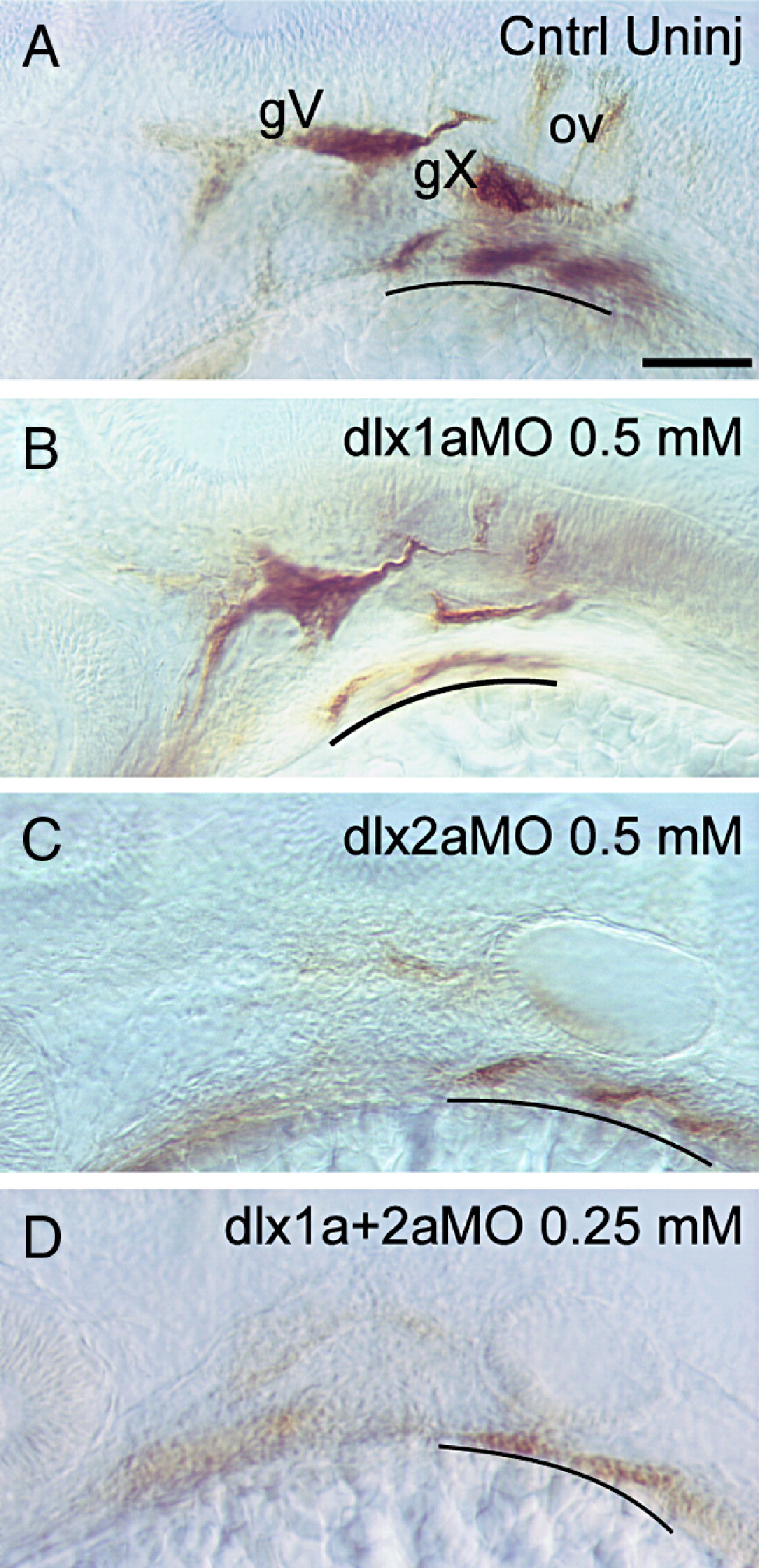
Fig. 7 Comparison of zn-5 staining in control and dlx1a, and dlx2a, and dlx1a/dlx2a morphants. Whole-mount immunohistochemical staining of 34 hpf embryos with the zn-5 antibody. Control embryos (A) are compared with embryos that were injected with (B) dlx1aMO, 0.5 mM; (C) dlx2aMO, 0.5 mM; (D) dlx1aMO + dlx2aMO 0.25 mM each. All panels are lateral views with anterior to the left. gV, trigeminal ganglia; gX, vagus ganglia; ov, otic vesicle; gill arches are underlined. Scale bar: 50 μm.
Figure Data
Acknowledgments
This image is the copyrighted work of the attributed author or publisher, and
ZFIN has permission only to display this image to its users.
Additional permissions should be obtained from the applicable author or publisher of the image.
Reprinted from Developmental Biology, 314(1), Sperber, S.M., Saxena, V., Hatch, G., and Ekker, M., Zebrafish dlx2a contributes to hindbrain neural crest survival, is necessary for differentiation of sensory ganglia and functions with dlx1a in maturation of the arch cartilage elements, 59-70, Copyright (2008) with permission from Elsevier. Full text @ Dev. Biol.