
|
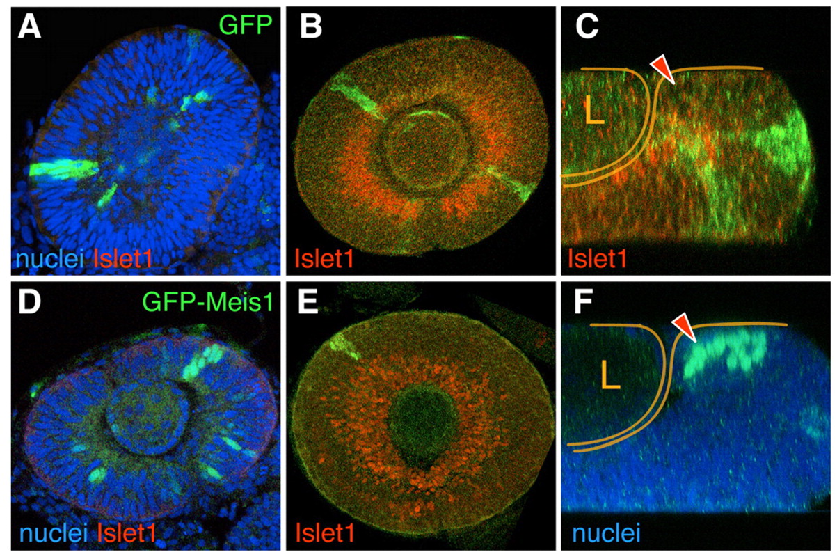
Fig. 4 Clonal overerexpression of meis1 in the developing eye prevents differentiation and results in cell sorting. Single optical sections from confocal z-stacks of GFP (A-C) or GFP-Meis1 (D-F) expressing clones induced genetically in developing eyes. GFP-meis1 signal is nuclear. At 24-30hpf, both clone types frequently span the whole width of the neuroepithelium (A,D). Confocal optical sections through the central retina (B,E) and z-sections (C,F) of 48hpf eyes. At this stage, GFP clones comprise both Islet1-expressing and non-expressing cells (B,C). By contrast, same-stage GFP-Meis1 clones in the central retina do not contain Islet1-positive cells (E). GFP-Meis1 clones are often located in the CMZ (F*ge, 9. The arrowheads (C,F) point to the CMZ, and the retina and the lens (L) are outlined.