
|
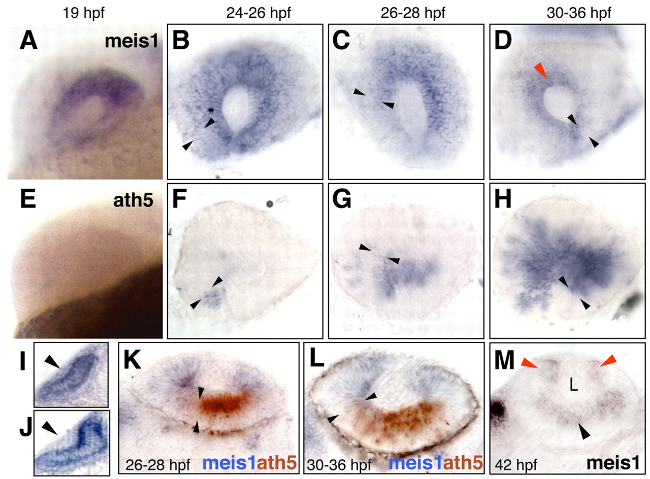
Fig. 1 meis1 retracts accompanying the ath5 wave and becomes restricted to the CMZ. (A-D) meis1 and (E-H) ath5 expression analyzed by single in situ hybridization. Developmental stages are indicated as hours post-fertilization (hpf) at 28.5°C. Lateral views of whole-mount (A,E) or dissected (B-D,F-H) eyes, with dorsal up and anterior to the left. The front of the ath5 domain is marked by black arrowheads. The red arrowhead in D points to meis1 expression in the prospective ciliary margin. (I-M) Transverse 40 μm vibratome sections. (I,J) Dorsal is up. meis1 is weakly expressed in the lens ectoderm before its thickening (I), but no signal is detected once the lens placode is formed (J). (K-M) Dorsal is to the left. meis1 and ath5 expression domains are complementary as shown by double in situ hybridization (K,L). Approximate limits of the ath5 signal are indicated by the black arrowheads. (M) At 42hpf, meis1 expression is detected by in situ hybridization in the ciliary margin (red arrowheads) and in the postmitotic ganglion cells (black arrowhead). L, lens.