
|
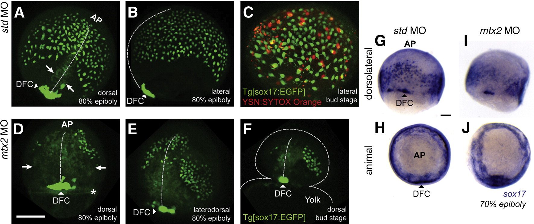
Fig. 6 Morphogenetic movements of mesendoderm are disrupted in mtx2 MO injected embryos. Live confocal imaging of std MO (A–C) and mtx2 MO (D–F) injected transgenic [sox17:EGFP+] embryos. std MO injected embryos at 80% epiboly (A, B) show the same expression pattern seen in sox17 in situs (G–J), with dorsal forerunner cells (DFC) showing the highest GFP+ expression (arrowheads in panels A and B). Axial mesoderm is not thought to express Sox17, but these cells are faintly visible (arrows) converging on the midline (dashed line) in panel A. (C) SYTOX-orange injection into the YSL of Tg[sox17:EGFP+] embryos shows Sox17:EGFP+ cells outnumber YSN by approximately 4:1. In mtx2 MO injected Tg[sox17:EGFP+] embryos, GFP+ cells are detected (D, E), but are disorganized and fail to descend vegetally. An unknown, circumferential ring-like structure was also visible in mtx2 MO injected embryos at this time point (asterC GFP+ cells are detected (D, E), but are disorganized and fail to descend vegetally. An unknown, circumferential riisk in panel D). Axial mesoderm cells in mtx2 MO injected embryos are broadly spread out (arrows in panel D) from the midline (vertical dashed line in panels D–F) even at the last stage before yolk lysis (F). (G–J) The sox17 expression pattern in std MO (G, H) and mtx2 MO (I, J) injected embryos at 60–70% epiboly parallel the GFP fluorescence pattern seen in the Tg[sox17:EGFP] line. Scale bar = 200 μm in panel D (applies to panels A–F), 100 μm in panel G (applies to panels G–J).
Reprinted from Developmental Biology, 314(1), Wilkins, S.J., Yoong, S., Verkade, H., Mizoguchi, T., Plowman, S.J., Hancock, J.F., Kikuchi, Y., Heath, J.K., and Perkins, A.C., Mtx2 directs zebrafish morphogenetic movements during epiboly by regulating microfilament formation, 12-22, Copyright (2008) with permission from Elsevier. Full text @ Dev. Biol.