
|
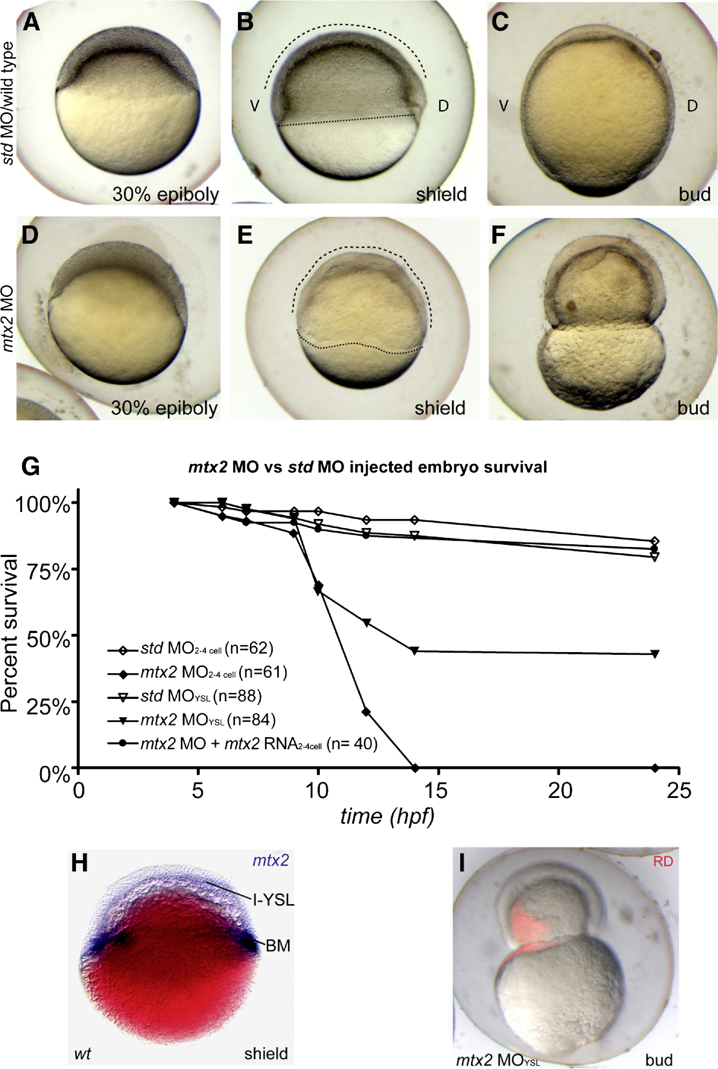
Fig. 1 Mtx2 is essential for epiboly. (A-F) Lateral views through epiboly, ″D″ and ″V″ mark dorsal and ventral sides of embryos, respectively. Std MO controls (A–C) proceeded through epiboly normally. However, mtx2 MO injected embryos displayed an epiboly defect phenotype (D-F). Specifically, mtx2 MO injected embryos appeared similar to controls until 30% epiboly. When controls reached shield stage (B), mtx2 MO injected embryos had an irregular blastoderm cap and blastoderm margin (dashed and dotted lines, respectively, in panels B and D) and had not clearly specified a shield. Lysis of mtx2 MO injected embryos occurred, due to blastoderm constriction (F), close to the timing of bud stage in std MO embryos (C). (G) Survival rates for embryos injected with std or mtx2 MO either early (two- to four-cell stage), late (YSL) or with 100 pg mtx2 capped RNA (two- to four-cell stage). (H) Shield stage whole-mount in situ for mtx2 in a wiFi> MO either early (two- to four-cell stage), late (YSL) or with 100 pg mtx2 capped RNA (two- to four-cell sld-type embryo, Nomarski optical section. The I-YSL and blastoderm margin (BM) staining pattern is indicated. (I) A merged bright/dark-field image of a bud stage embryo injected at 1000 stage with mtx2 MO and rhodamine dextran (RD).
Reprinted from Developmental Biology, 314(1), Wilkins, S.J., Yoong, S., Verkade, H., Mizoguchi, T., Plowman, S.J., Hancock, J.F., Kikuchi, Y., Heath, J.K., and Perkins, A.C., Mtx2 directs zebrafish morphogenetic movements during epiboly by regulating microfilament formation, 12-22, Copyright (2008) with permission from Elsevier. Full text @ Dev. Biol.