
Image
|
Figure Caption
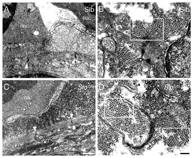
Fig. S3 Heart muscle ultrastructure is unaffected by the sloθ45 mutation. Transverse TEM sections through the heart tube at 48 hpf reveal a thin myofibrillar band in the heart wall in both siblings (A) and slou45 mutants (C). Z-disc to Z-disc distances show no obvious differences between the mutant and sibling (arrowheads, A and C). Longitudinal sections reveal some myofibrils in cross section (boxes, B and D) and in these cases it is clear that the myofibrils contain both thick and thin filaments. RBC, red blood cell. Scale bars: 500 nm in A,C; 200 nm in B,D.
Figure Data
Acknowledgments
This image is the copyrighted work of the attributed author or publisher, and
ZFIN has permission only to display this image to its users.
Additional permissions should be obtained from the applicable author or publisher of the image.
Full text @ Development