
|
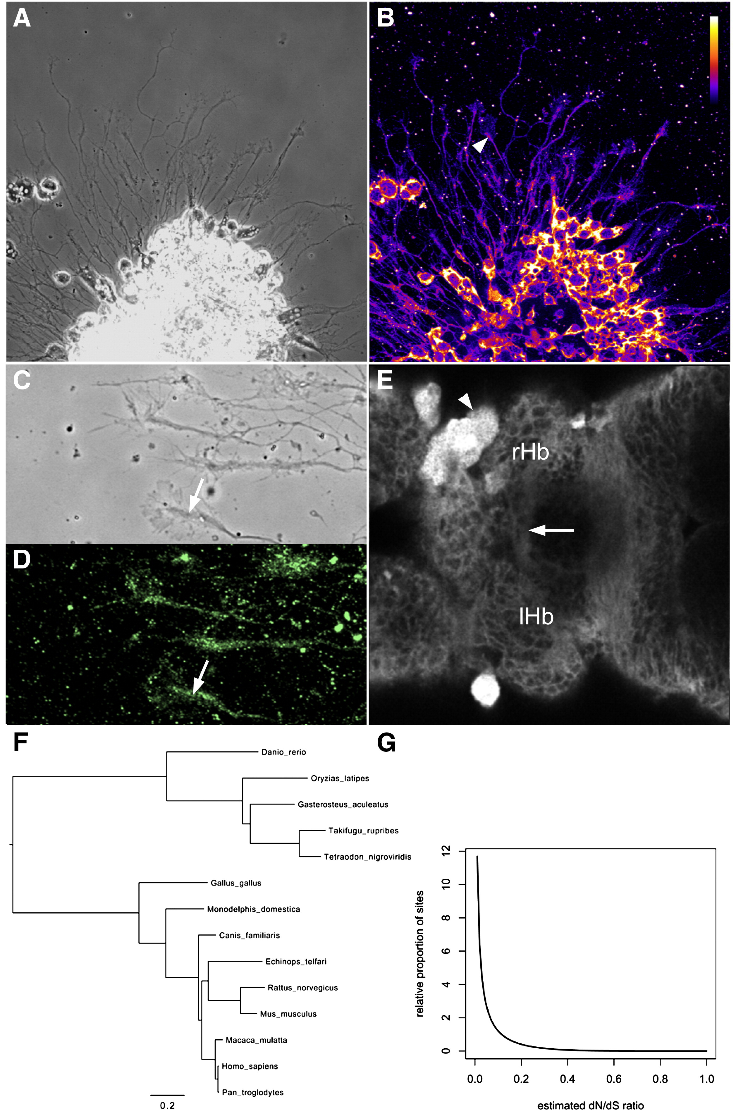
Fig. 4 Esrom is a broadly expressed and slowly evolving protein. (A, B) Axons extending from cultured forebrain neurons. The 7014 antibody to Esrom labels all cell bodies and axons. The core (arrowhead) of growth cones is more strongly labeled than the periphery. (C, D) Cultured axons labeled with the 2a anti-PAM antibody, with strong label in the phase-dense region (arrows). (E) An optical section through a 3 dpf brain labeled with the 7014 antibody. Axons in the habenular commissure are indicated (arrow). A xanthophore (arrowhead) is brightly labeled, both in the cytoplasm and nucleus. (F) The phylogeny used for the analysis of codon evolution. (G) The estimated distribution of evolutionary rate variation across codon sites (measured as the dN/dS ratio). The vast majority of sites have very low dN/dS ratios, suggesting that most codon sites are subject to very strong negative (stabilizing) selection.
Reprinted from Molecular and cellular neurosciences, 37(2), Hendricks, M., Mathuru, A.S., Wang, H., Silander, O., Kee, M.Z., and Jesuthasan, S., Disruption of Esrom and Ryk identifies the roof plate boundary as an intermediate target for commissure formation, 271-283, Copyright (2008) with permission from Elsevier. Full text @ Mol. Cell Neurosci.