
|
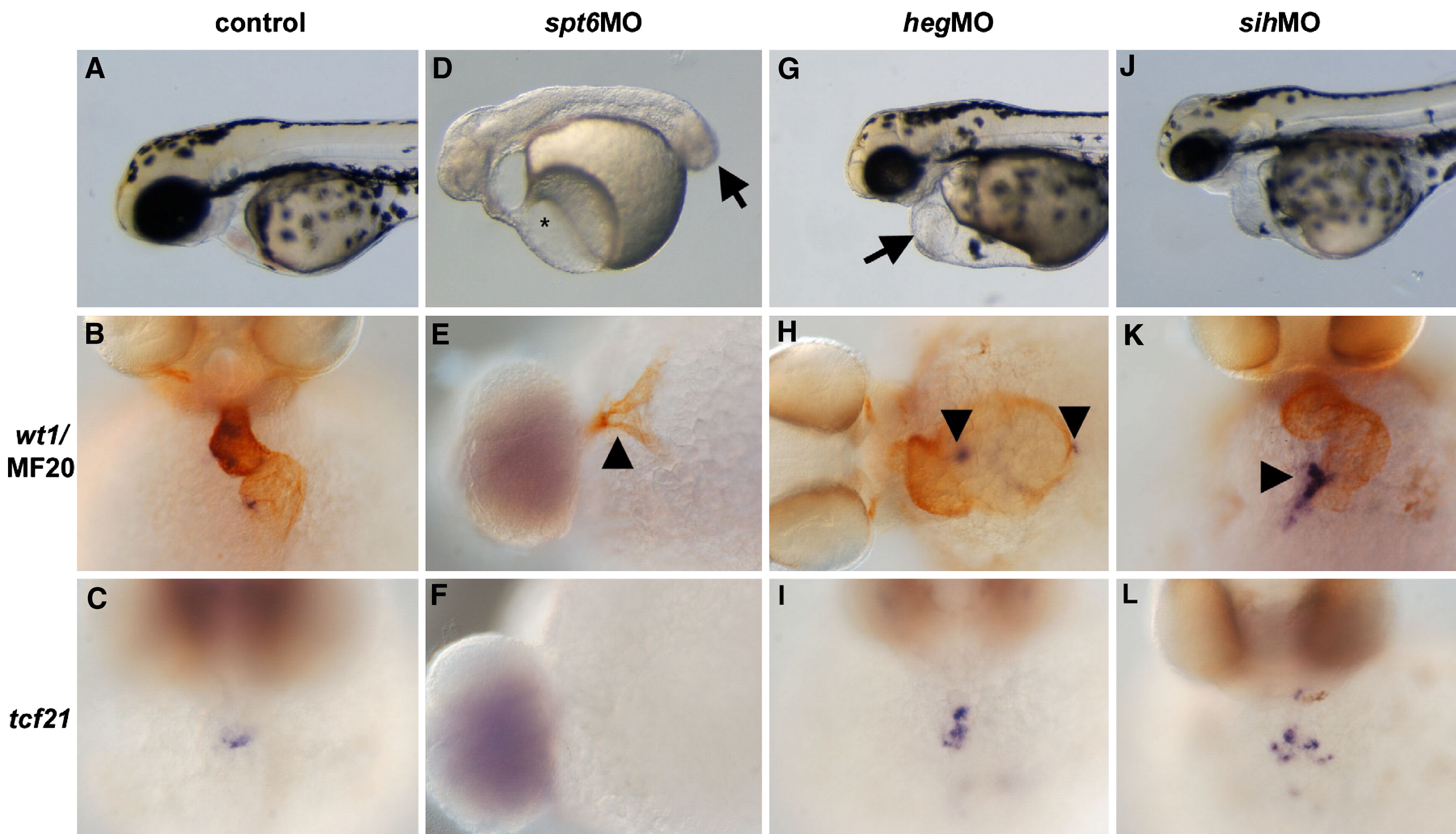
Fig. 6 pandora/spt6 is required for the PEO lineage. (A) Buffer-injected control embryos shown as a live 48 hpf embryo, (B) stained with wt1 (blue)/MF20 (brown) to mark the PEO and myocardium respectively, and with tcf21 (C) as a second marker of the PEO. (D) spt6MO embryos shown as live 48 hpf embryos. Note the extensive pericardial edema (asterisk) and tail extension defects (arrow). The PEO is not visible either by (E) wt1 or (F) tcf21 staining. Myocardium is still present, however, as evidenced by the MF20 positive tissue in the wt1/MF20 double-stained spt6MO embryos (arrowhead in panel E). (G) hegMO embryos display an enlarged heart (arrow) but relatively a relatively normal PEO as assayed by (H) wt1, with two wt1-positive clusters at the characteristic positions (arrowheads) and (I) tcf21 expression. (J) sihMO embryos shown live at 48 hpf. Staining with (K) wt1 and (L) >wt1-positive clusters at the characteristic positions (arrowheads) and (I) tcf21 expression. (J) sihMO embryos shown live at 48 hpf. Staining with (K) wt1 and (L) tcf21 reveals no effect of heart function on the formation of the PEO (arrowhead in panel K). Anterior is to the left in all panels except B, C, I, K and L which are face views with dorsal at the top of the panel.
Reprinted from Developmental Biology, 315(1), Serluca, F.C., Development of the proepicardial organ in the zebrafish, 18-27, Copyright (2008) with permission from Elsevier. Full text @ Dev. Biol.