
|
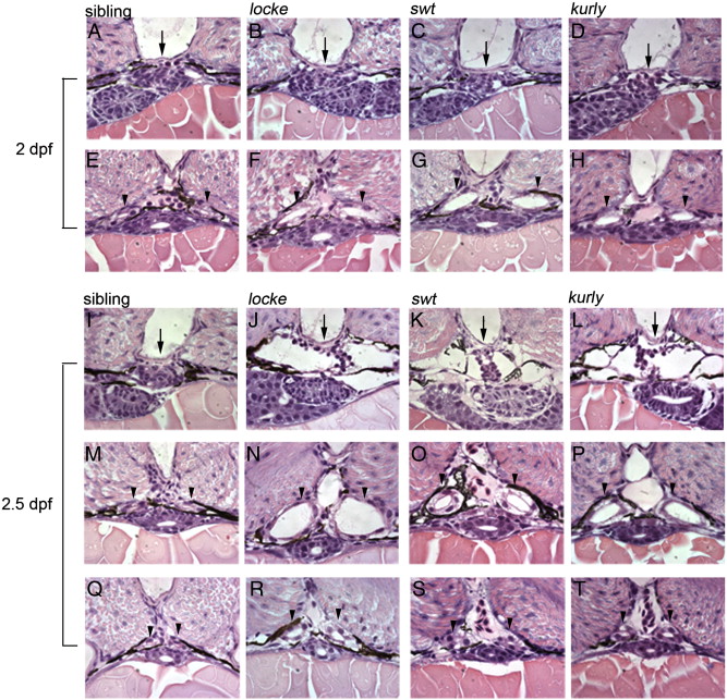
Fig. 3 Temporal and spatial analysis of cystic kidneys in zebrafish mutants. (A–H) 2 dpf; (I–T) 2.5 dpf. At 2 dpf, the glomerulus (arrows, A–D) appears intact in the mutant embryos; however, the medial tubules have become dilated (arrowheads, E–H). At 2.5 dpf, the glomerulus (arrows, I–L) and surrounding Bowman's space have become enlarged, compressing the podocytes in the mutants. The medial tubules are grossly dilated (arrowheads, M–P). Interestingly, the posterior tubules are less dilated, approaching wild-type size (arrowheads, Q–T). All pictures are 4 μm JB-4 plastic sections, stained with hematoxylin and eosin and taken with a 100x oil objective lens.
Reprinted from Developmental Biology, 314(2), Sullivan-Brown, J., Schottenfeld, J., Okabe, N., Hostetter, C.L., Serluca, F.C., Thiberge, S.Y., and Burdine, R.D., Zebrafish mutations affecting cilia motility share similar cystic phenotypes and suggest a mechanism of cyst formation that differs from pkd2 morphants, 261-275, Copyright (2008) with permission from Elsevier. Full text @ Dev. Biol.