
|
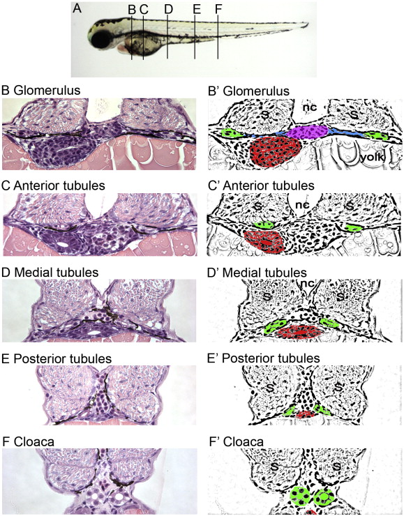
Fig. 2 Histological and schematic representations of the pronephros along the anterior to posterior axis. (A) Depiction of the corresponding regions referred to in the text. (B, C, D, E, F) Histological sections stained with hematoxylin and eosin. Pictures taken with a 40x objective lens. (B′, C′, D′, E′, F′) Schematic diagrams highlighting regions of interest. (B, B′) Glomerulus: The glomerulus (pink) is found ventral to the notochord (nc) and medial to either somite (s). Connecting to the glomerulus are tubules that extend laterally (blue). The tubules then turn at the edge of the somites and extend toward the posterior (green). Also shown is the gut (red). (C, C′) Anterior tubules: The region designated as the anterior tubules is slightly posterior to the glomerulus region in which the viscera can be observed. (D, D′) Medial tubules: In this region, the gut (red) and tubules (green) are positioned toward the midline and ventral to the notochord (nc). (E, E′) Posterior tubules: The gut has become smaller and the tubules (green) are positioned more medially. (F, F′) Cloaca: This is the most posterior section before the tubules fuse into a single opening outside the body.
Reprinted from Developmental Biology, 314(2), Sullivan-Brown, J., Schottenfeld, J., Okabe, N., Hostetter, C.L., Serluca, F.C., Thiberge, S.Y., and Burdine, R.D., Zebrafish mutations affecting cilia motility share similar cystic phenotypes and suggest a mechanism of cyst formation that differs from pkd2 morphants, 261-275, Copyright (2008) with permission from Elsevier. Full text @ Dev. Biol.