
Image
|
Figure Caption
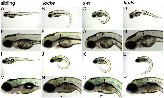
Fig. 1 locke, swt and kurly mutants develop pronephric cysts. (A–H) 3 dpf, (I–P) 5 dpf. At 3 dpf, locke, swt and kurly mutants are easily identified by their “curly-tail down” phenotype. Panels E–H are higher magnification images of panels A–D, showing the cystic dilations visible under light microscopy. At 5 dpf, the cystic dilations have increased in size shown in panels I–L and magnified in panels M–P. The black arrowheads mark the location of the cysts, posterior to the eye and ear. In general, locke mutants have smaller cysts than swt or kurly.
Figure Data
Acknowledgments
This image is the copyrighted work of the attributed author or publisher, and
ZFIN has permission only to display this image to its users.
Additional permissions should be obtained from the applicable author or publisher of the image.
Reprinted from Developmental Biology, 314(2), Sullivan-Brown, J., Schottenfeld, J., Okabe, N., Hostetter, C.L., Serluca, F.C., Thiberge, S.Y., and Burdine, R.D., Zebrafish mutations affecting cilia motility share similar cystic phenotypes and suggest a mechanism of cyst formation that differs from pkd2 morphants, 261-275, Copyright (2008) with permission from Elsevier. Full text @ Dev. Biol.