
|
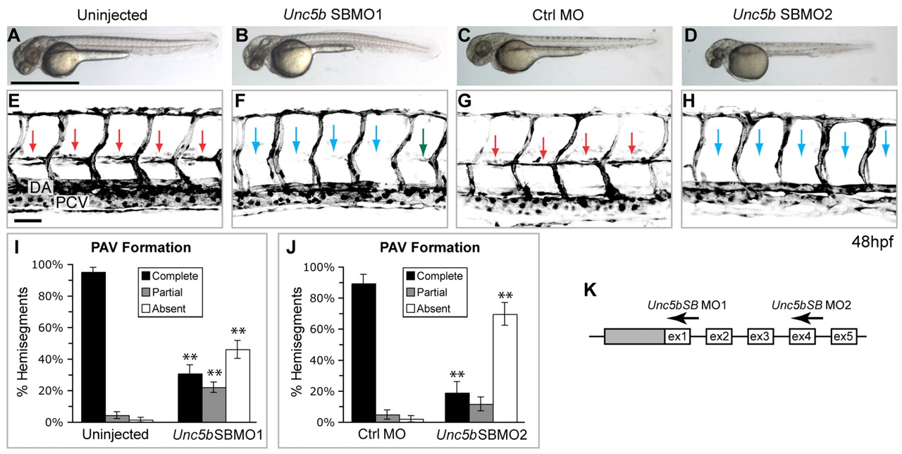
Fig. 4 Knockdown of zebrafish unc5b prevents formation of a specific vessel: the PAV. (A-D) Bright-field images of live embryos, anterior towards the left. Overall trunk morphology is normal in unc5b morphants (1 ng unc5bSBMO1 or 4 ng unc5bSBMO2) compared with controls (uninjected or 8 ng control MO). (E-H) Confocal projections of fli:egfp transgenics (reverse contrast) show that unc5b knockdown prevents PAV formation. Lateral views of somites 7-11, anterior towards the left; confocal projections through entire trunk. Embryos imaged live (G,H) or fixed after anti-GFP staining (E,F). (E,G) In uninjected embryos or control morphants at 48 hpf, almost every hemisegment is spanned by a PAV at the horizontal myoseptum (red arrows). (F,H) In embryos injected with either of two unc5b splice-blocking MOs, PAVs are absent (blue arrows) or only partially span their hemisegment (green arrows). DA, dorsal aorta; PCV, posterior cardinal vein. (I,J) PAV formation scored as percentage of hemisegments per embryo. In controls, at least 98% of hemisegments have complete or partial PAVs; this fraction is drastically reduced by either unc5b MO. n=25-30 embryos per condition; **P<0.0001. Bars show mean±s.e.m. (K) Partial unc5b genomic structure (not to scale), showing 5′ UTR (gray box), coding exons (white boxes) and MO targets. Scale bars: 1 mm in A for A-D; 50 μm in E for E-H.