
|
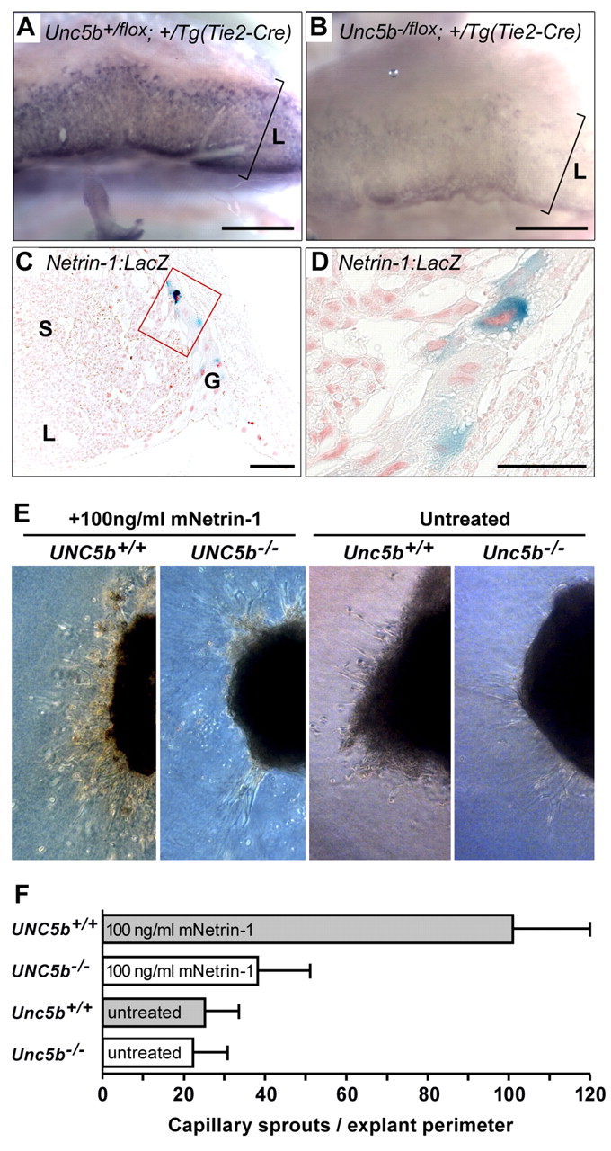
Fig. 3 Unc5b/Netrin signaling in the placenta. (A,B) Unc5b mRNA localization in the labyrinth. Placentas from E12.5 embryos were hemisected and probed with antisense RNA complementary to Unc5b mRNA. (A) Unc5b+/flox;+/Tg(Tie2-Cre) and (B) Unc5b-/flox; +/Tg(Tie2-Cre); L indicates extent of labyrinthine layer. Scale bars: 1 mm. (C,D) Netrin expression in giant trophoblast cells. Placentas (E12.5) from embryos heterozygous for the netrin1:lacZ gene trap (Serafini et al., 1996) were fixed, hemisected and stained with X-gal to detect lacZ activity; 10 μm sections were mounted on slides and counterstained with nuclear Fast Red. L, labyrinth; S, spongiotrophoblast; G, giant trophoblast cell layer. Image in D is a 4x magnification of boxed area in C. Scale bars: 200 μm in C; 100 μm in D. (E) Growth of umbilical vessels in vitro. Umbilical vessels at E10.5 were explanted into collagen gels and allowed to sprout for 10 days in the presence or absence of netrin 1 protein. Numbers of sprouts were counted under phase contrast. Genotypes and growth conditions are indicated above each frame. (F) Quantitation from four pairs of umbilical arteries of each genotype. Error bars are standard deviation.