
|
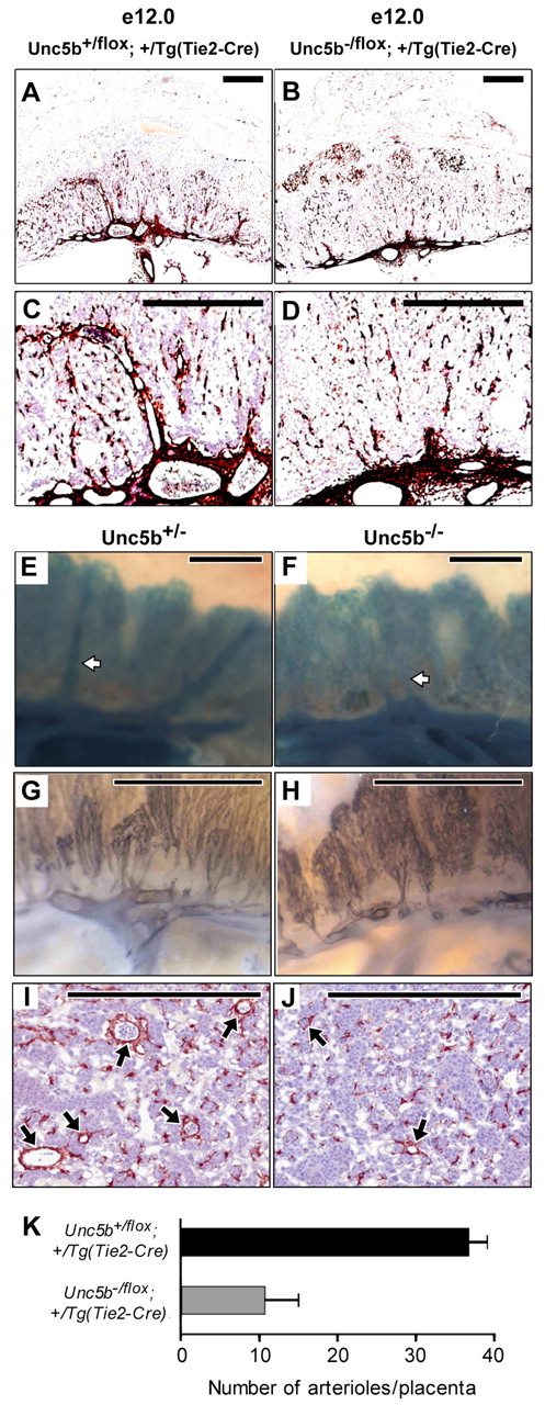
Fig. 1 Vascular ablation of Unc5b impacts labyrinthine arterioles. Embryos were harvested at E12.0 from Unc5bflox/flox x Unc5b+/-; Tg(Tie2-Cre)/Tg(Tie2-Cre) (A-D), Unc5b+/-;R26R [Rosa26 Reporter (Soriano, 1999)] x Unc5b+/-; Tg(Tie2-Cre)/Tg(Tie2-Cre) (E,F) or Unc5b+/- x Unc5b+/- (G-J) matings. Placentas were dissected, formalin fixed, hemisected and stained for: smooth muscle actin (anti-smooth muscle actin antibody followed by HRP-conjugated secondary antibody) to identify smooth muscle cells surrounding the arterial bed (A-D); for β-galactosidase activity (X-gal) to identify endothelial expression of Tie2-Cre (E,F); and for Pecam l (anti-Pecam1 antibody followed by HRP-conjugated secondary antibody) to highlight the endothelium (G,H). Ta;-galactosidase activity (X-gal) to identify endothelial expression of Tie2-Cre (E,F); and for Pecam l (anti-Pecam1here is a decrease in the number of robust vertical vessel stalks in placentas lacking UNC5B (B,D,F,H) when compared with their littermate controls (A,C,E,G). This is also shown in I,J, which represent 10 μm cross-sections through samples stained for smooth muscle actin. Arterioles are indicated by arrows; scale bars: 0.5 mm. (K) The number of arterioles per placenta was determined by serial sectioning (10 μm) of hemisected placentas, staining for smooth muscle actin and visually counting all vessels. Paired littermates (three pairs) representing each of the two genotypes were used for all measurements. Error bars are standard deviation.