
Image
|
Figure Caption
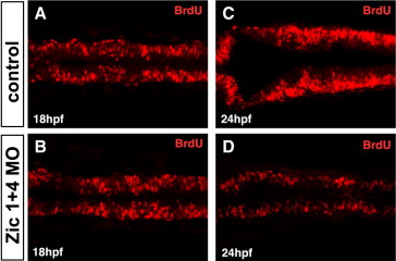
Fig. S2 Zic1 and Zic4 function is required after 18 hpf for dorsal neural proliferation in the hindbrain. (A–D) Dorsal confocal sections (3 μm thick; longitudinal view) through hindbrain immunolabeled with BrdU, marking cells in the S-phase of the cell cycle, at 18 hpf (A,B) and 24 hpf (C,D) in control (A,C) and Zic1 + 4 morphants (B,D). No change in hindbrain proliferation is detected at 18 hpf in Zic1 + 4 morphants (B, n = 11) compared to control embryos (A, n = 10), whereas at 24 hpf, proliferation is markedly reduced in Zic1 + 4 morphants (D, n = 8) compared to control embryos (C, n = 13).
Figure Data
Acknowledgments
This image is the copyrighted work of the attributed author or publisher, and
ZFIN has permission only to display this image to its users.
Additional permissions should be obtained from the applicable author or publisher of the image.
Reprinted from Developmental Biology, 314(2), Elsen, G.E., Choi, L.Y., Millen, K.J., Grinblat, Y., and Prince, V.E., Zic1 and Zic4 regulate zebrafish roof plate specification and hindbrain ventricle morphogenesis, 376-392, Copyright (2008) with permission from Elsevier. Full text @ Dev. Biol.