
|
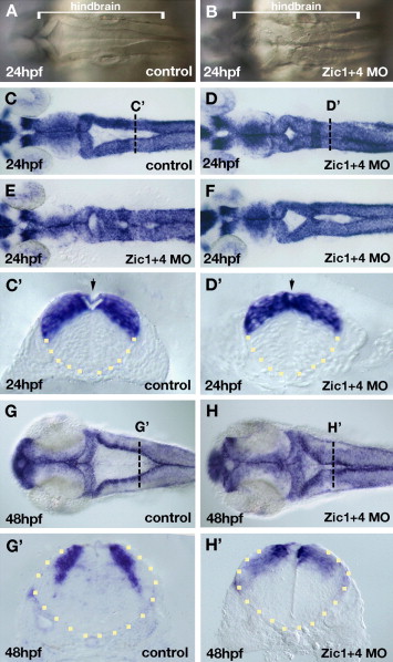
Fig. 2 Dorsal hindbrain ventricle morphogenesis is altered in Zic1 and Zic4 morphants. Dorsal view of the hindbrain (A–F, G, H) and cross-sections of the neural tube (outlined by yellow dots) at the level of r5 (C′, D′, G′, H′). (A, B) Bright field live images of hindbrains of 24 hpf control embryos (A) and Zic1 + 4 morphants (B). Dorsal folds that outline the hindbrain ventricle in control hindbrains (A) are aberrantly fused at the dorsal midline in Zic1 + 4 morphants (B). Brackets in panels A and B indicate the length of the hindbrain. (C–F) Dorsal zic1 expression at 24 hpf allows visualization of hindbrain ventricle defects in Zic1 + 4 morphants (D–F; and see Table 1) compared to controls (C). Transverse sections at AP levels indicated by white dotted lines in panels C and D show hindbrain ventricle opening is impaired in 24 hpf Zic1 + 4 morphants (D′) compared to controls (C′). Arrow in panel C′ indicates roof plate (RP) overlying the ventricle, and in panel D′ indicates absence of a well-defined RP. (G–H′) Dorsal view of zic1 expression in 48 hpf controls (G) and Zic1 + 4 morphants (H) showing “dorsal fused” phenotype. (G′, H′) Transverse sections at AP levels indicated by the white dotted lines in panels G and H show ventricle opening is impaired in 48 hpf Zic1 + 4 morphants (H′) compared to controls (G′).
Reprinted from Developmental Biology, 314(2), Elsen, G.E., Choi, L.Y., Millen, K.J., Grinblat, Y., and Prince, V.E., Zic1 and Zic4 regulate zebrafish roof plate specification and hindbrain ventricle morphogenesis, 376-392, Copyright (2008) with permission from Elsevier. Full text @ Dev. Biol.