
|
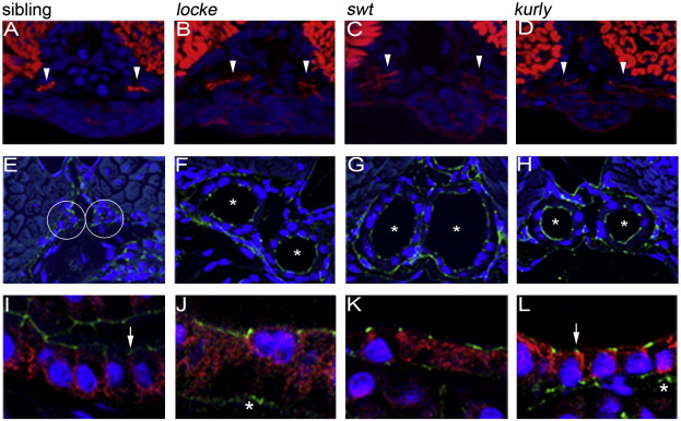
Fig. 6 Na+/K+ATPase localization is disrupted in cystic tissues, while apical polarity is maintained. Apical localization of F-actin (red) observed in 2 dpf wild-type siblings (A) is maintained in the medial tubules (white arrowheads) of mutants (B–D). The somites and gut are also stained by phalloidin (red). Correct apical localization of ZO-1 (green) is observed in 4 dpf wild-type sibling (E) and mutant (F–H) embryos. Sibling tubules are outlined by a white circle, and mutant tubules are distinguished by asterisks. Basolateral localization of the Na+/K+ATPase (red) observed in wild-type siblings (I) is altered in cystic mutants at 4 dpf (J–L). Note in panels J–L that Na+/K+ATPase is adjacent to apical ZO-1 expression (green; indicated by white arrows) which is not observed in wild-type (I). All confocal images were taken with a 40× water objective lens. Panels A–D are cryosections; panels E–L are plastic sections. Nuclei (blue) are stained with either DRAQ5 (A–D) or Hoechst (E–L). ZO-1 staining basal to the pronephros comes from a different tissue (asterisks in panels K and L) and does not reflect an alteration in ZO-1 within the pronephros.
Reprinted from Developmental Biology, 314(2), Sullivan-Brown, J., Schottenfeld, J., Okabe, N., Hostetter, C.L., Serluca, F.C., Thiberge, S.Y., and Burdine, R.D., Zebrafish mutations affecting cilia motility share similar cystic phenotypes and suggest a mechanism of cyst formation that differs from pkd2 morphants, 261-275, Copyright (2008) with permission from Elsevier. Full text @ Dev. Biol.