
|
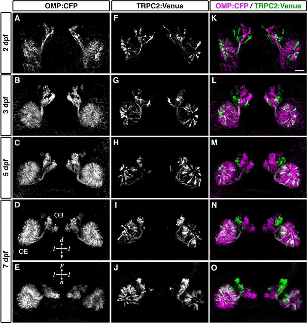
Fig. 7 Establishment of the segregated axonal terminations at early stages of development. Time-lapse imaging of labeled OSNs in living OMP:CFP;TRPC2:Venus zebrafish embryos was performed. CFP-labeled (A-E; magenta in K-O) and Venus-labeled OSNs (F-J; green in K-O) in a single representative embryo are shown. All photographs represent stacked images of optical sections. Axonal terminations from CFP-labeled and Venus-labeled OSNs were already segregated at 2 dpf (K) and maintained through development (L, 3 dpf; M, 5 dpf; N, O, 7 dpf). A-D, F-I, K-N, Frontal views; E, J, O, dorsal views. l, Lateral; d, dorsal; v, ventral; p, posterior; a, anterior. Scale bar, 50 μm.