
|
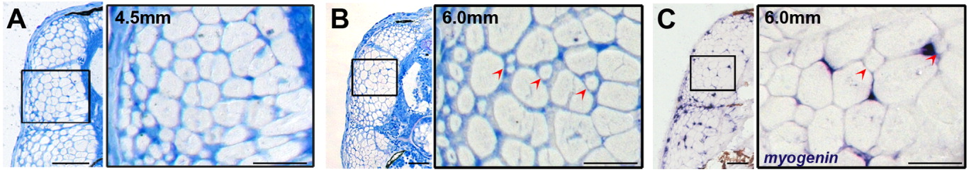
Fig. 1 Hyperplastic growth of trunk muscle in zebrafish larvae. A: Methylene blue-stained section through the trunk of a 4.5 mm larva. Hyperplastic growth at this stage is predominantly by stratified hyperplasia. Note the abundance of small diameter fibers at the lateral surface of the fast muscle, and absence of small diameter fibers within the fast muscle. B: Methylene blue-stained section through the trunk of a 6.0 mm larva. Small fibers are apparent within the fast muscle, indicating mosaic hyperplasia (red arrowheads) C: Myogenin labeling in a 6.0 mm larval trunk. Note the mosaic appearance of myogenin labeling in the trunk, suggesting mosaic differentiation of muscle. Mosaic hyperplasia is also evident by the appearance of small diameter fibers within the fast muscle (red arrowheads). Scale bars = 50 μm in A-C, 25 μm in insets.