
|
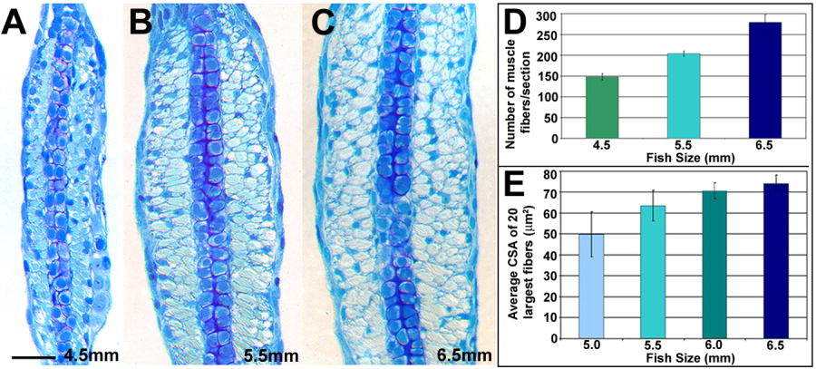
Fig. 4 Growth of muscle in the late larval pectoral fin occurs by hyperplasia and hypertrophy. A-C: Transverse sections of methylene blue-stained pectoral fins during the late larval period. There is a large increase in overall size of the pectoral fin musculature from 4.5 mm (A) to 6.5 mm (C). D: Quantification of total muscle fibers/section shows an increase in the number of muscle fibers during the late larval period. Between 4.5 and 6.5 mm, the number of fibers almost doubles, increasing from a mean of 148 fibers to a mean of 279 fibers (4.5 mm, n = 3; 5.5 mm, n = 4; 6.5 mm, n = 4). D: Increase in the mean cross-sectional area of the 20 largest fibers suggests that hypertrophy is contributing to muscle growth during this period (all sizes, n = 3). Scale bar = 25 μm in A-C.