
|
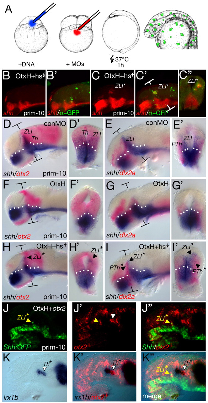
Fig. 5 Otx2 rescues the ZLI in OtxH embryos. Gene expression analysis of embryos at prim-10 (28 hpf); the markers are indicated on each panel. (A) Embryos were injected with 60 pg of the heat-shock-inducible otx2-GFP DNA construct into the one-cell-stage blastomere. Embryos were kept at 28°C until reaching the four-cell stage. Then, the double morpholino (MO) mix was injected to block transcription of endogenous otx1l and otx2. At the one-somite stage, the embryos were heat-shocked for 1 hour at 37°C and were fixed at the prim-12 stage. To reliably localise the Otx2-GFP-positive clone, an FITC-coupled antibody staining against GFP and a Fast Red single in situ hybridisation for shh were performed. Injection of otx2-GFP DNA and heat-shock activation led to a random induction of GFP-positive clones in the OtxH embryos. Confocal microscopy analysis of the embryos showed that ectopic induction of shh expression is generally not observed in otx2-GFP clones (B,B′). However, when these clones were located within the MDT, shh expression was induced in these cells (C,C′ and cross-section in C″, 13/16-4/6 detected via &alpha-GFP staining and 9/10 via GFP-ISH). (D,D′) To mark the prethalamic-thalamic boundary, a double in situ hybridisation of otx2 and shh was performed. (E,E′) To mark the prethalamus, a double staining with dlx2a and shh was performed. (F-G′) In control (con) embryos, shh expression is absent at the ZLI. In addition, the otx2 expression domain is smaller (F,F′) and the dlx2a domain is absent (G,G′). By contrast, in embryos in which otx2-GFP is heat-activated, shh expression can be rescued in the presumptive MDT (H,H′), and a cross-section reveals that the induction is independent of the basal shh expression domain (H′). The rescue of shh in small clones is sufficient to induce the expression of dlx2a (5/10; I); for example, its expression was induced uni-laterally in the neural tube (I′). T-shaped brackets mark the plane of the section in the following picture. The dotted line marks the border between the alar and basal plate. (J-K″) otx2 mRNA (150 pg; + red-lineage tracer) was injected into one of 64 cells of OtxH shh-GFP embryos. At 32 hpf, embryos were fixed and stained for GFP (green). The provision of an ectopic otx2 message is able to rescue Shh-GFP expression within the ZLI in OtxH embryos (J-J″, yellow arrowhead), as shown by a co-localisation of the red-lineage tracer of the Otx2+ cells with the green GFP expression. The irx1b expression profile was assessed in the same embryo (K-K″). When Otx2+ cells are located within the presumptive thalamic territory, they express irx1b (white arrowhead), shown by co-localisation of the red lineage tracer with irx1b detection (blue). (K″) Merged picture of Shh-GFP expression, the red lineage tracer and irx1b expression. prim-10, primordium stage 10; PTh, prethalamus; PTh*, rescued prethalamus; Th, thalamus; ZLI, zona limitans intrathalamica; ZLI*, rescued zona limitans intrathalamica.