
|
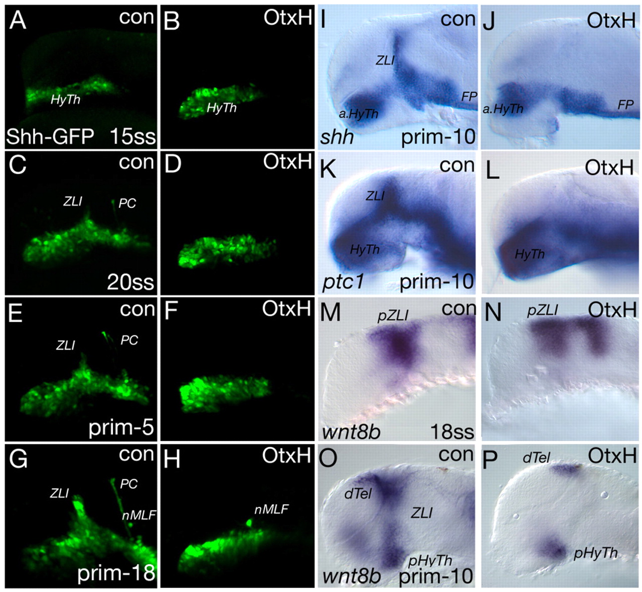
Fig. 3 Analysis of the ZLI in OtxH embryos. Confocal microscopy analysis of transgenic shh-GFP OtxH embryos and controls (con) (A-H) and gene expression analysis of OtxH embryos (I-P); the markers and stages are indicated. (A,B) Shh-GFP is detectable in the hypothalamic primordium (HyTh) in control embryos as well as in OtxH embryos at 17.5 hpf. (C-H) From 21 to 32 hpf, the ZLI develops from ventral to dorsal and the posterior commissure (PC) forms at the diencephalic-mesencephalic boundary in control embryos (C,E,G), whereas, in OtxH embryos, neither the ZLI nor the PC is detectable (D,F,H). Interestingly, the nucleus of the medio-longitudinal fascicle (nMLF) is visible in control embryos as well as in OtxH embryos (G,H). (I,J) At prim-10 (28 hpf), the Shh expression domains of the basal plate are unaltered in the anterior hypothalamus (aHyTh) and floor plate (FP) in OtxH embryos, whereas the ZLI, located in the alar plate, is missing compared with control embryos (42/51). (K,L) Similarly, the expression domain of ptc1 is absent at the ZLI in OtxH embryos (31/48). (M,N) Expression of the marker of the presumptive ZLI (pZLI), wnt8b, is unaltered in the diencephalic expression domain at 18 somites (18 hpf), although the MHB expression shifts anteriorly. (O,P) At prim-10, wnt8b is absent in the ZLI (10/12) but persists in the dorsal telencephalon (dTel) and posterior hypothalamus (pHyTh). prim, primordium stage; ZLI, zona limitans intrathalamica.