
|
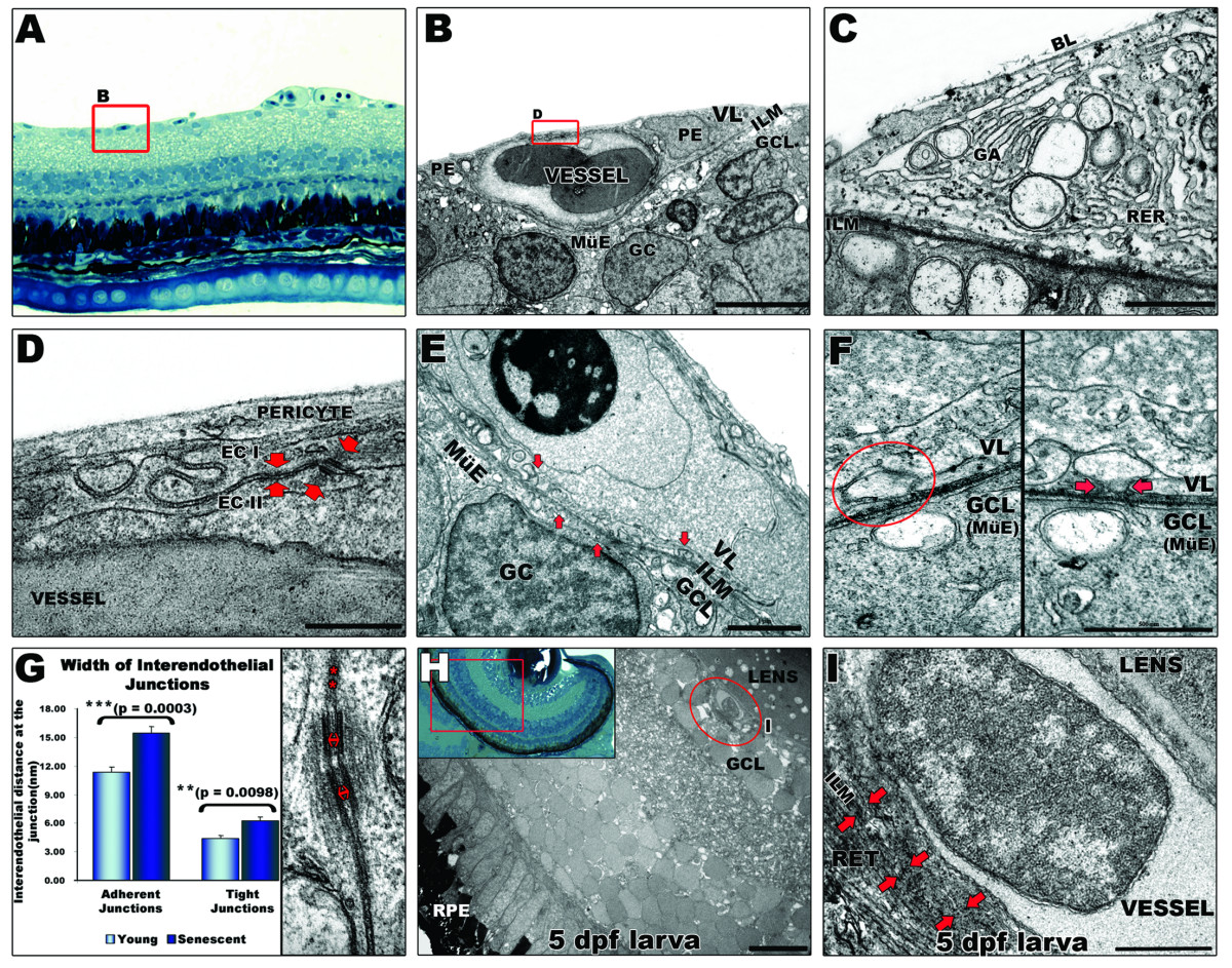
Fig. 3 Ultrastructural analysis of the inner retina blood supply in zebrafish. A: Light microscopy image of blood vessels (highlighted by a red square) overlying the inner limiting membrane (ILM) of the retina in adult zebrafish. 40× magnification. B: Ultrastructure of capillary. The basal lamina (BL) is in contact with the ILM enclosing pericytes (PE) and the vascular endothelium. Scale bar: 5 μm. C: Typical structural features of pericytes, e. g. Golgi apparatus (GA), rough endoplasmic reticulum (RER) and large membrane bound vesicles. Scale bar: 1 μm. D: Vitreal space lined by pericyte overlying endothelial cells (EC) which display interdigitating junctional complexes (arrows). Scale bar: 500 nm. E: Multiple vesicles (arrows) from 20 to 250 nm contacting the inner limiting membrane of the retina indicate active interaction between the vessels and the ganglion cell layer (at the Müller endfeet). Scale bar: 1 μm. F: Vascular (top) and ganglion cell layer interface (bottom). A vesicle (left panel) apparently separated from the cell membrane, fuses with the cell membrane when the section is tilted by 43° (right panel) suggesting transcellular transport. Scale bar: 500 nm. G: Interendothelial junctions are significantly more open in the senescent fish. Distance between endothelial cells at tight (asterisks) and adherent (arrows) junctions were measured in different peripheral and central areas of retinas from senescent and young adult fish. Error bars: sem. Inset in H: Histology of a 5 dpf zebrafish eye. 40× magnification. H: EM image of a 5 dpf larval eye illustrating the relationship of hyaloid vessels (encircled by a red line) to both the lens and the retina. Scale bar: 10 μm. I: Higher magnification of the hyaloid vessel confirms its tight attachment to the lens and looser contact to the retina (arrows). Scale bar: 1 μm. MüE: Müller cell endfeet; GC: ganglion cell; GCL: ganglion cell layer; VL: vascular layer; RPE: retinal pigmented epithelium; RET: retina.