
Image
|
Figure Caption
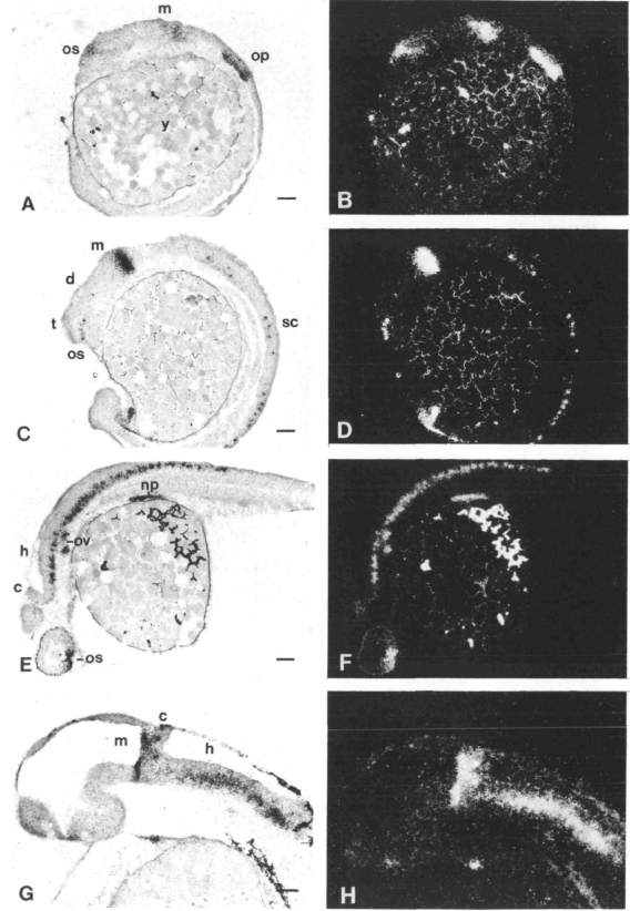
Fig. 5 Localization of pax[zf-b] transcripts by in situ hybridization in tissue sections of zebrafish embryos at different developmental stages. Sagittal sections are shown for embryos after 12 h (A,B), 18 h (C,D), 24 h (E,F) and 36 h (G,H) of development. Bright-field and dark-field images of each section are shown side by side. The embryos are oriented with their anterior end to the left. Abbreviations: c, cerebellum; d, diencephalon; h, hindbrain; m, midbrain; np, nephritic primordium; op, otic placode; os, optic stalk; ov, otic vesicle; t, telencephalon; y, yolk. Bars, 50 μm.
Figure Data
Acknowledgments
This image is the copyrighted work of the attributed author or publisher, and
ZFIN has permission only to display this image to its users.
Additional permissions should be obtained from the applicable author or publisher of the image.
Full text @ Development